





Waxaa soo dhigay: Northwest Eye gudaha Guud ahaan on April 30, 2026
Hordhac
Many people feel uncertain about how to care for their eyes, yet understanding this vital organ can truly change how we see the world. By learning about the eye’s structure, you can take meaningful steps towards protecting your vision and overall health. It’s common to feel unsure about what affects your eyesight. Let’s explore how the different parts of your eyes work together to help you see clearly and stay healthy.
Define Eyeball Anatomy: Key Components and Structure
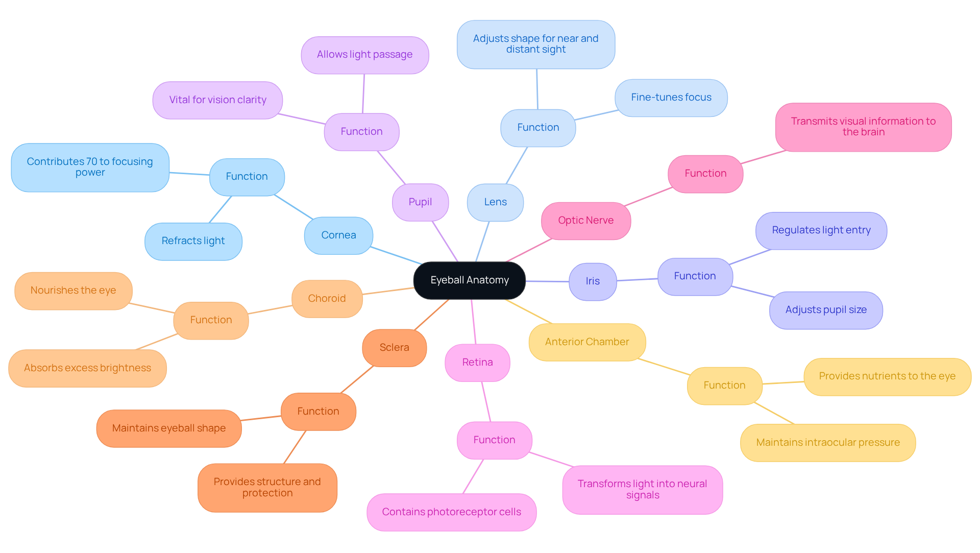
We understand that navigating the complexities of eye health can feel overwhelming, especially when considering procedures like qalliinka cataract. The anatomy of the eyeball is a remarkable organ, composed of several vital components that work together to enable sight. Let’s take a closer look at these essential structures:
- Cornea: This transparent front layer refracts light entering the eye, contributing approximately 70% to the eye’s focusing power.
- Muraayadaha: A flexible structure that fine-tunes the focus of rays onto the retina, adjusting its shape for both near and distant sight. This is crucial for optimal visual outcomes, especially for those undergoing cataract surgery.
- Iris: The colored section of the eye that adjusts the size of the pupil, regulating how much light enters.
- Arday: The aperture in the center of the iris that allows light to pass through, playing a vital role in vision clarity.
- Retina: The innermost layer containing photoreceptor cells (rods and cones) that transform light into neural signals, essential for visual perception.
- Nerfaha Indhaha: This nerve transmits visual information from the retina to the brain, enabling us to interpret images.
- Sclera: The white outer layer that provides structure and protection to the eyeball, maintaining its shape.
- Choroid: A layer rich in blood vessels that nourishes the eye and absorbs excess brightness, supporting overall eye health.
- Anterior Chamber: The space filled with aqueous humor located between the cornea and the iris, which helps maintain intraocular pressure and provides nutrients to the eye.
We know that understanding how your eyes work is essential for feeling confident about your vision and health. This knowledge is particularly important for individuals undergoing procedures like cataract surgery. At Northwest Eye, we’re here to help you navigate the costs of eye care, offering flexible financing options to ease your journey. Our goal is to ensure that you can receive the necessary treatments without financial strain. As David Turbert observes, “By assisting in directing rays as they enter the eye, the cornea and the lens both play crucial roles in providing us with clear sight.” By understanding the anatomy of the eyeball, you can take proactive steps towards maintaining your vision and overall eye health.
Contextualize Eyeball Anatomy: Importance for Vision and Health
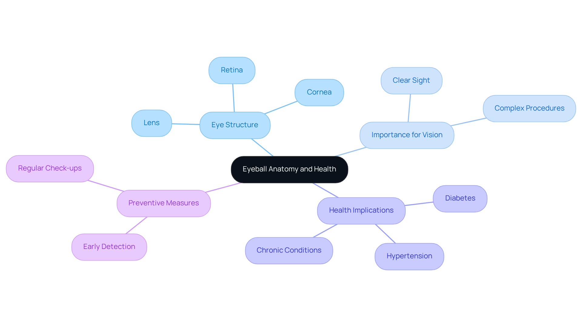
We understand that the structure of your eyes can feel overwhelming, but it’s essential for your sight and overall health. Each part works together to help you see clearly, which is something we all cherish. This complex procedure is crucial for aragti cad.
It’s concerning to think that many people may not be aware of their risk for vision loss, and we want to help you stay informed and proactive. The health of these ocular structures is closely linked to various systemic diseases. For instance, changes in the retina can serve as early indicators of conditions such as diabetes and hypertension. Baaritaannada joogtada ah ee indhaha can facilitate early detection of systemic diseases, which is vital for your health.
Understanding how your eyes work can empower you to take steps to protect your vision and health. Regular check-ups not only help in preserving eyesight but also in identifying potential health issues before they escalate. Effective interventions for eye conditions, including early detection and timely treatment, can significantly improve outcomes for patients, particularly those with chronic conditions like diabetic retinopathy, which affects millions globally.
By prioritizing your caafimaadka indhaha, you’re taking a vital step towards safeguarding your overall well-being.
Trace the Evolution of Eyeball Anatomy: Historical Perspectives
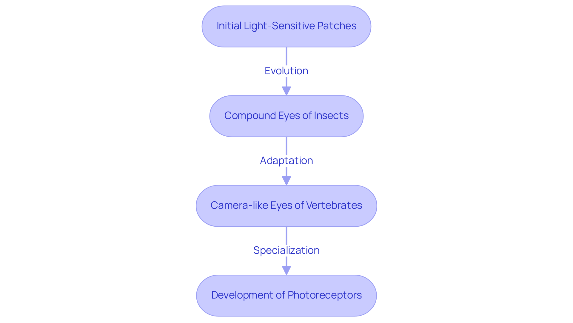
We understand that the evolution of the anatomy of the eyeball can seem complex, but it’s a fascinating journey of adaptation that reflects the ingenuity of nature. Initially, the simplest visual organs were just light-sensitive patches in early life forms. Over time, these evolved into intricate systems capable of creating images. For example, the compound eyes of insects represent an early adaptation that provides a wide field of sight. In contrast, vertebrates developed camera-like eyes, and the anatomy of the eyeball includes lenses that provide better focus and clarity. Interestingly, research shows that the vertebrate eye evolved from non-visual organs around 420 million years ago, marking a significant transition that speaks to the resilience of life.
Throughout evolutionary history, significant transformations in the anatomy of the eyeball have resulted in the sophisticated eye structures we see today. The creation of a light-sensitive layer oo leh specialized photoreceptors has been vital in improving how we see the world, allowing for sharper images and vibrant colors. Specialists like Karl Deisseroth have explored the development of these photoreceptors, emphasizing their role in enhancing sight. Moreover, the eye has emerged through convergent evolution as many as 40 times or more, showcasing the adaptability of eye structures across various species. This remarkable adaptability not only showcases the eye’s functionality but also inspires us to appreciate the wonders of evolution in our own lives.
Detail Key Characteristics of Eyeball Anatomy: Functions and Parts
We understand that taking care of your eyes can feel overwhelming, but understanding the anatomy of the eyeball and how each part contributes to your vision can empower you to take charge of your eye health.
Each component of the eyeball plays a crucial role in vision:
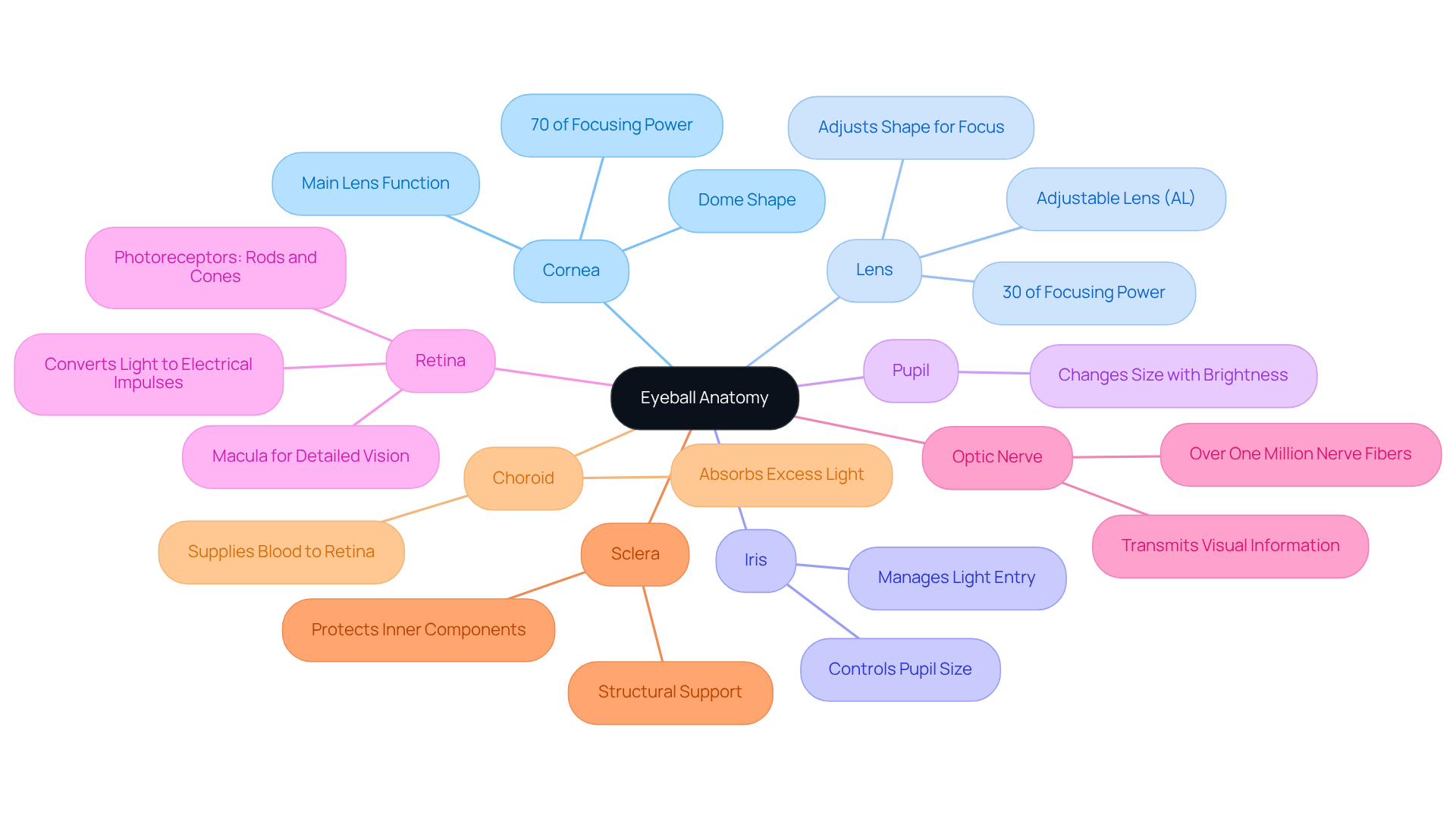
- Cornea: Did you know that your cornea acts like the main lens of your eye? In the anatomy of the eyeball, this transparent layer bends light to focus it onto the retina, providing about 70% of the eye’s focusing power due to its dome shape.
- Muraayadaha: You might be surprised to learn that in the anatomy of the eyeball, your lens changes shape to help you see things clearly, no matter how far away they are. The anatomy of the eyeball, located just behind the iris, adjusts to concentrate rays from objects at varying distances, contributing about 30% of the eye’s focusing power. At Northwest Eye, we offer the innovative Adjustable Lens (AL), which can be modified after surgery using UV rays to enhance sight based on your lifestyle preferences.
- Iris: The colored section of your eye is part of the anatomy of the eyeball, and it adjusts the size of the pupil, managing how much light enters your eye. Understanding the anatomy of the eyeball is crucial for optimal perception in different lighting conditions.
- Arday: This opening changes size in response to brightness levels, helping you see clearly and comfortably.
- Retina: The anatomy of the eyeball includes light-sensitive tissue located at the back of your eye, which contains photoreceptors-rods for low-light conditions and cones for color perception. As part of the anatomy of the eyeball, it converts light signals into electrical impulses sent to your brain for processing. The anatomy of the eyeball includes the macula, which has a high concentration of photoreceptors essential for detailed vision.
- Nerfaha Indhaha: As part of the anatomy of the eyeball, the optic nerve comprises over one million nerve fibers that transmit visual information from the light-sensitive layer of your eye to your brain, enabling you to perceive images.
- Sclera: The tough, white outer layer of the eye, crucial to the anatomy of the eyeball, provides structural support and protection for the inner components.
- Choroid: This vascular layer, which is important in the anatomy of the eyeball, supplies blood to the retina and absorbs excess light, preventing reflection within your eye.
By understanding the anatomy of the eyeball and how each part functions, you can better appreciate the importance of eye health and its impact on your daily life. Baaritaannada joogtada ah ee indhaha can be a simple yet powerful way to safeguard your vision and overall well-being.
Gunaanad
We understand that understanding the anatomy of the eyeball can feel overwhelming, but it’s essential for appreciating how it influences your vision and overall health. Every part of your eye, from the cornea to the retina, works together to help you see the world around you, showing just how amazing this organ truly is. When you understand how these parts work, you can feel more confident in making choices about your eye care and health.
Taking care of your eyes is about more than just seeing well; it’s a crucial part of feeling good overall. Regular eye examinations can help catch issues early, leading to better health outcomes. By taking these steps, you’re not just caring for your eyes; you’re investing in your overall well-being and future health.
Su'aalaha Badiya La Weydiiyo
What are the main components of eyeball anatomy?
The main components of eyeball anatomy include the cornea, lens, iris, pupil, retina, optic nerve, sclera, choroid, and anterior chamber.
Waa maxay shaqada cornea-ga?
The cornea is the transparent front layer of the eye that refracts light entering the eye, contributing approximately 70% to the eye’s focusing power.
Sidee buu muraayaddu uga qayb qaadataa aragga?
The lens is a flexible structure that fine-tunes the focus of light rays onto the retina, adjusting its shape for both near and distant sight, which is crucial for optimal visual outcomes.
What role does the iris play in vision?
The iris is the colored section of the eye that adjusts the size of the pupil, regulating how much light enters the eye.
Waa maxay ujeeddada ardayga?
The pupil is the aperture in the center of the iris that allows light to pass through, playing a vital role in vision clarity.
What does the retina do?
The retina is the innermost layer of the eye containing photoreceptor cells (rods and cones) that transform light into neural signals, essential for visual perception.
How does the optic nerve function?
The optic nerve transmits visual information from the retina to the brain, enabling us to interpret images.
What is the role of the sclera?
The sclera is the white outer layer of the eye that provides structure and protection, maintaining the shape of the eyeball.
What is the choroid and its function?
The choroid is a layer rich in blood vessels that nourishes the eye and absorbs excess brightness, supporting overall eye health.
What is the anterior chamber?
The anterior chamber is the space filled with aqueous humor located between the cornea and the iris, which helps maintain intraocular pressure and provides nutrients to the eye.
Liiska Ilaha
- Define Eyeball Anatomy: Key Components and Structure
- eyewiki.org (https://eyewiki.org/Eye_in_Numbers)
- 19 Oraah oo Ku Saabsan Aragtida oo Ka Dhigi Doonta Inaad Nolosha Si Ka Duwan U Aragto | Aragtida Ka Baxsan (https://beyondvision.ca/2015/11/19-quotes-about-vision-that-will-make-you-see-life-differently)
- Eye Anatomy: Parts of the Eye and How We See (https://aao.org/eye-health/anatomy/parts-of-eye)
- Contextualize Eyeball Anatomy: Importance for Vision and Health
- Xarumaha Xakamaynta iyo Kahortagga Cudurrada (https://cdc.gov/vision-health/data-research/vision-loss-facts)
- World report on vision (https://who.int/publications-detail-redirect/world-report-on-vision)
- Aragga oo daciif ah iyo indho la'aan (https://who.int/news-room/fact-sheets/detail/blindness-and-visual-impairment)
- Tirakoobka Caafimaadka Indhaha - Akadeemiyada Mareykanka ee Cilmi-baarista Indhaha (https://aao.org/newsroom/eye-health-statistics)
- Trace the Evolution of Eyeball Anatomy: Historical Perspectives
- scientificamerican.com (https://scientificamerican.com/article/evolution-of-the-eye)
- answersingenesis.org (https://answersingenesis.org/human-body/eyes/darwins-challenge?srsltid=AfmBOoqAW_4J5aBBlFXVYBXDb36s3dy_PLD9CWAg_up4nyZpZdL7gXmf)
- manchester.ac.uk (https://manchester.ac.uk/about/news/443-million-year-old-fossils-reveal-early-vertebrate-eyes)
- Detail Key Characteristics of Eyeball Anatomy: Functions and Parts
- Eye Anatomy: Parts of the Eye and How We See (https://aao.org/eye-health/anatomy/parts-of-eye)
- contactsdirect.com (https://contactsdirect.com/parts-of-eye)
- Eyes: A Window to the World for Your Brain (https://my.clevelandclinic.org/health/body/21823-eyes)
- eyewiki.org (https://eyewiki.org/Eye_in_Numbers)
- Anatomy of the Eye (https://hopkinsmedicine.org/health/conditions-and-diseases/anatomy-of-the-eye)