Posted by: Northwest Eye in General on May 17, 2025
Overview
Retinal detachment is a serious condition that can be alarming. It occurs when the light-sensitive membrane at the back of the eye separates from its supportive tissue. If not treated promptly, it can lead to significant vision loss. We understand how concerning this can be for you.
Recognizing early symptoms, such as flashes of light or increased floaters, is crucial. By understanding the types, causes, and available treatment options, you can take proactive steps toward effective intervention. It’s common to feel overwhelmed, but early action can make a difference in preserving your eyesight.
Remember, we are here to help you through this process. Seeking care and support is essential, and you don’t have to face this alone. Your vision matters, and taking these steps can lead to a positive outcome.
Introduction
Retinal detachment is a critical eye condition that can lead to significant vision loss if not promptly addressed. We understand how concerning this can be. This serious issue occurs when the retina, a vital layer of tissue at the back of the eye, separates from its supportive structure, disrupting the conversion of light into neural signals. Understanding the intricacies of retinal detachment—from its various types and causes to the symptoms that serve as warning signs—is essential for early detection and treatment.
With approximately 1 in 10,000 people affected each year, awareness and education about this condition can empower individuals to seek timely medical intervention. It’s common to feel anxious about what this means for your vision and quality of life. As the landscape of treatment continues to evolve, exploring both surgical and non-surgical options reveals a promising outlook for those at risk. We are here to help you through this process and ensure you receive the care you need.
Define Retinal Detachment: Understanding the Basics
The serious condition of retinal detachment can understandably cause concern. It occurs when the light-sensitive membrane, a delicate layer of tissue at the back of the eye, separates from its supportive tissue. If not addressed promptly, retinal detachment can result in significant loss of sight. The eye’s inner layer is essential for converting light into neural signals, which the brain uses for visual recognition. When retinal detachment occurs, the retina’s ability to function is compromised, often resulting in blurred vision or even blindness. While blurred vision can stem from various issues like nearsightedness, farsightedness, and astigmatism, it may also indicate eye diseases such as cataracts or diabetic retinopathy. If left untreated, these symptoms can lead to serious health complications, so it is vital to seek professional medical help immediately if you experience blurred vision.
There are three primary types of retinal detachment:
- Rhegmatogenous Detachment: This is the most common type, caused by a tear or break in the eye’s inner layer, allowing fluid to seep underneath and separate it from the underlying tissue.
- Tractional Detachment: This occurs when scar tissue on the surface of the eye layer pulls it away from the underlying tissue, often seen in patients with diabetes.
- Exudative Detachment: This type is characterized by fluid accumulation beneath the retina without any tears or breaks, typically due to inflammatory conditions or tumors.
Recent data suggests that about 1 in 10,000 individuals encounter separation of the eye’s inner layer each year. While this condition is infrequent, its impact on sight can be considerable. Experts emphasize the importance of early diagnosis and treatment of retinal detachment, as timely intervention can greatly improve patient outcomes. For instance, a study on treatment results for eye separation showed that surgical action can restore sight in most cases, especially when performed within the initial days of symptom emergence.
Understanding the fundamentals of eye separation is crucial for identifying its signs, which may include sudden flashes of light, floaters, or a shadow over the visual field. Recognizing these indicators can lead to quicker medical assistance, ultimately protecting your eyesight and enhancing your quality of life. As prominent eye specialists have noted, early identification and intervention for retinal detachment are essential for maintaining sight and avoiding permanent harm. If you experience blurred vision, particularly if related to cataracts, it is vital to seek immediate medical attention. Incorporating insights from case studies on treatment outcomes can further illustrate the effectiveness of timely surgical intervention, reinforcing the importance of seeking immediate care when symptoms arise.
Explore Causes of Retinal Detachment: Risk Factors and Mechanisms
Various factors can contribute to the risk of retinal detachment, which is a serious condition.
- Aging: We understand that as we age, the natural process can lead to the vitreous gel within the eye shrinking and pulling away from the retina, which may result in tears.
- Eye Injury: It’s common to feel concerned about trauma to the eye, as it is a major factor that can result in tears or holes in the retina, elevating the risk of separation. Data show that around 20% of eye separations are due to eye injuries, emphasizing the significance of protective measures.
- Previous Eye Surgery: If you’ve had surgical procedures, especially cataract operations, you may worry about an increased risk of separation of the retina due to alterations in the eye’s structure.
- High Myopia: Individuals with severe nearsightedness might feel more vulnerable, as the elongation of the eyeball can predispose them to eye problems.
- Family History: A genetic inclination can also contribute, making certain individuals more susceptible to eye separation.
Understanding these risk factors is crucial for all of us, particularly because aging populations are especially vulnerable, and the risk of retinal detachment rises as we grow older. Identifying these factors can empower you to seek prompt medical guidance, particularly if you encounter symptoms like flashes or floaters in your sight. As Amir Khan, M.D. states, “Most eye floaters don’t require treatment, but your eye doctor likely will recommend regular eye exams to ensure the condition doesn’t worsen.”
We encourage you to consult with an ophthalmologist promptly to prevent lasting eyesight loss, as early detection and treatment can greatly enhance outcomes. This urgency is highlighted by the case study titled ‘Retinal Detachment Awareness,’ which emphasizes that prompt consultation is vital for preventing retinal detachment and maintaining sight.
Identify Symptoms of Retinal Detachment: Early Warning Signs
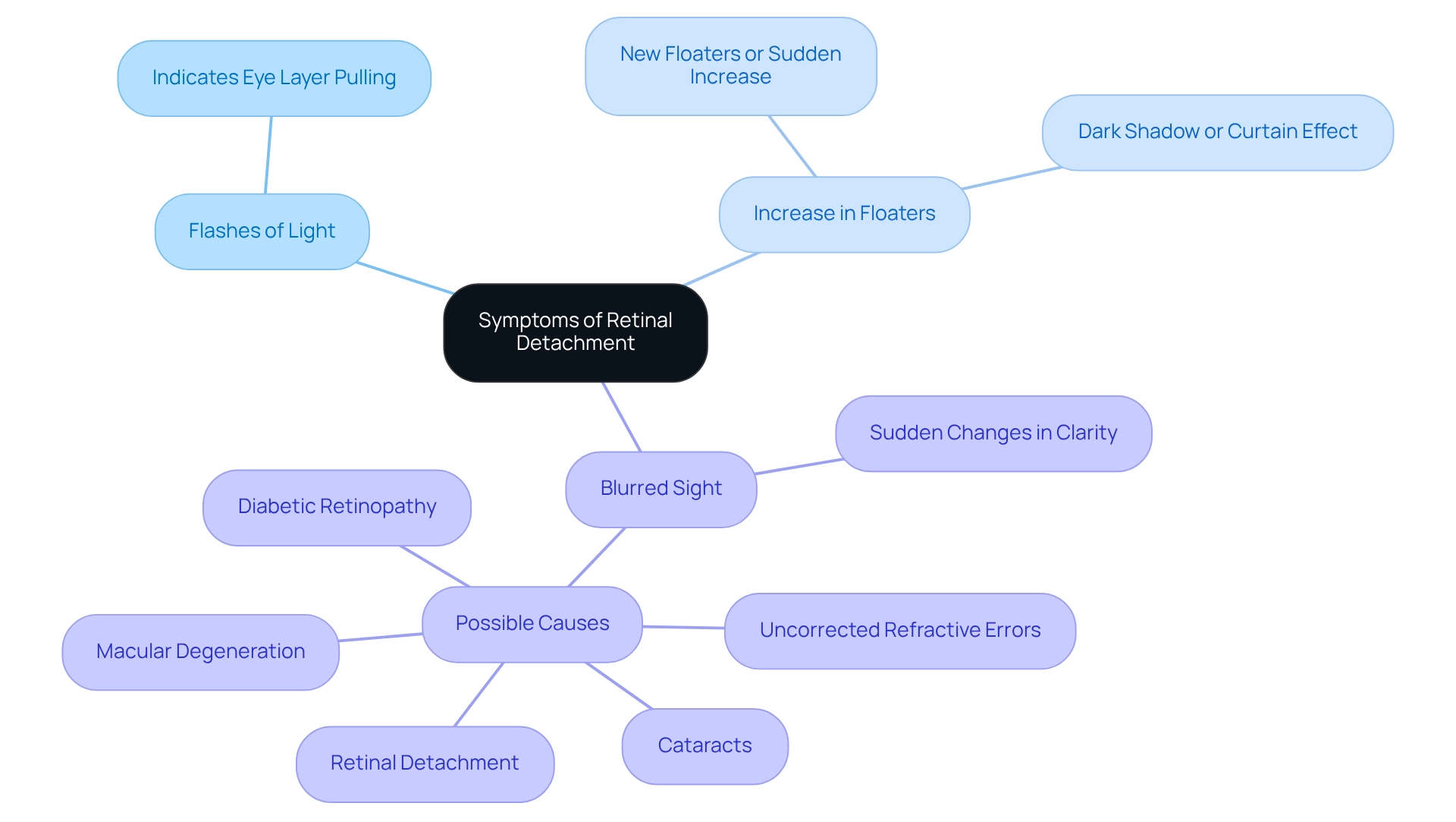
We understand that it’s important to be aware of the common symptoms of retinal detachment, as they can be concerning. Here are some key signs to watch for:
- Flashes of Light: You might notice sudden flashes, which can indicate that the eye’s light-sensitive layer is being pulled.
- Increase in Floaters: The appearance of new floaters or a sudden increase in existing floaters could signal a problem that needs attention. If you see a dark shadow or curtain-like effect in your peripheral vision, it may suggest that you are experiencing retinal detachment.
- Blurred Sight: Sudden changes in clarity can also be a warning sign. Blurred vision can arise from various conditions, including uncorrected refractive errors, cataracts, diabetic retinopathy, macular degeneration, or even retinal detachment. If you experience any of these symptoms, it is crucial to seek immediate medical attention to prevent further damage. We are here to help you through this process. If you are experiencing blurred vision or any other concerning symptoms, please request a consultation now.

Understand Diagnosis of Retinal Detachment: Procedures and Techniques
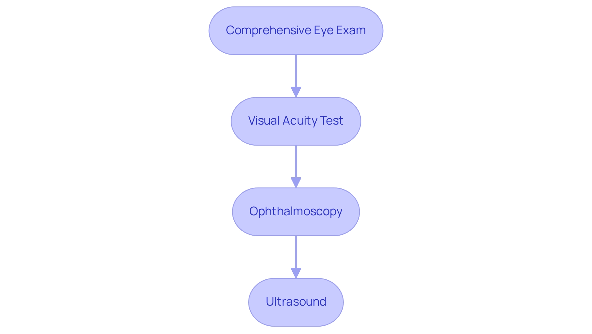
We understand that the possibility of retinal detachment can be concerning. Diagnosing this condition typically involves several important steps that are designed to ensure your comfort and clarity throughout the process.
- Comprehensive Eye Exam: An eye care professional will perform a thorough examination, often using dilating drops to widen your pupil for better visibility. This step is crucial for a complete assessment of your eye health.
- Visual Acuity Test: This test assesses how well you can see at various distances, helping us understand your specific situation more clearly.
- Ophthalmoscopy: A special instrument is used to examine the back of your eye for any tears or separation. This allows us to identify any potential issues directly.
- Ultrasound: In cases where the view of the retina is obstructed, ultrasound imaging can help visualize the retina. This technology is vital for accurate diagnosis of retinal detachment, and these diagnostic procedures are essential for confirming the presence of retinal detachment and determining the appropriate treatment. We are here to help you through this process, ensuring you feel supported every step of the way.

Review Treatment Options for Retinal Detachment: Surgical and Non-Surgical Approaches
Caring for eye separation encompasses a variety of surgical and non-surgical methods tailored to the specific type and severity of the separation. We understand that timely intervention is crucial for preserving vision, as delays can lead to irreversible damage. Here are some key treatment options that may offer you hope:
- Laser Surgery (Photocoagulation): This technique uses lasers to seal retinal tears, effectively preventing further detachment. It is often utilized for early-stage separations and has shown high success rates, providing reassurance for many patients.
- Cryopexy: This method employs extreme cold to create scar tissue that secures the light-sensitive layer in place, offering a reliable solution for certain types of separations.
- Scleral Buckle: In this surgical procedure, a silicone band is placed around the eye, gently pushing the eye wall against the retina. This technique has a strong track record of success, especially for more severe retinal detachment cases. Statistics indicate that scleral buckle procedures yield favorable results, with many patients experiencing significant improvements in their vision.
- Vitrectomy: This procedure involves removing the vitreous gel that may be exerting pressure on the light-sensitive tissue at the back of the eye, often accompanied by the repair of any related tears. It is a common approach for complex cases, and many find comfort in its effectiveness.
- Pneumatic Retinopexy: In this innovative technique, a gas bubble is injected into the eye to assist in reattaching the light-sensitive layer affected by retinal detachment. Recent advancements have led to minimally invasive methods, allowing patients to return to their normal activities as soon as the next day.
Expert opinions highlight the evolution of these techniques. Dr. Rajeev Muni notes that new in-office methods may enable patients to have their retina attached without incisions or strict positioning, significantly enhancing recovery times. He emphasizes that this is a new approach where patients might be able to return to all their usual activities almost immediately. Furthermore, ten years of study has focused on improving eye reattachment methods to enhance patients’ visual quality.
Real-world examples underscore the effectiveness of laser surgery, with many patients enjoying successful outcomes and rapid recovery. The case study titled ‘Minimally Invasive Technique for Retinal Detachment Repair’ illustrates how these new techniques enhance recovery times for patients suffering from retinal detachment and minimize complications associated with traditional surgical methods. Overall, the landscape of eye separation treatment continues to evolve, offering patients a variety of effective options to protect their vision. We are here to help you through this process and support you every step of the way.
Implement Prevention Strategies for Retinal Detachment: Protecting Your Vision
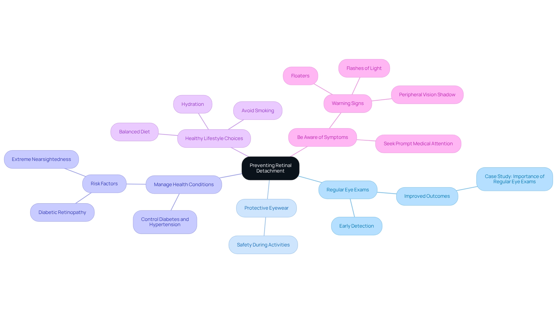
To effectively reduce the risk of retinal detachment, we encourage you to consider implementing the following strategies:
- Regular Eye Exams: We understand how crucial it is to remain vigilant about your eye health. Routine comprehensive dilated eye exams are essential for the early detection of retinal issues, including small tears that could lead to retinal detachment. Studies show that regular check-ups significantly enhance the chances of timely intervention and successful treatment. For instance, a case study highlights that early detection through regular exams can lead to significantly improved outcomes for patients.
- Protective Eyewear: It’s common to overlook safety during activities that present a risk of eye injury. Using safety glasses can help avert damage that may result in separation of the retina.
- Manage Health Conditions: We know that keeping conditions such as diabetes and hypertension under control is crucial. These can elevate the risk of eye complications, including separation. Statistics suggest that diabetic retinopathy and severe nearsightedness are major risk factors for the separation of the retina.
- Healthy Lifestyle Choices: A balanced diet abundant in antioxidants, sufficient hydration, and steering clear of smoking can enhance your overall eye health. These choices can assist in reducing risks linked to retinal detachment.
- Be Aware of Symptoms: Educating yourself about the warning signs of retinal detachment—such as sudden flashes of light, floaters, or a shadow in your peripheral vision—enables you to seek prompt medical attention, which is vital for preserving vision. It’s important to note that if there is a retinal detachment, a second treatment or surgery may be necessary.
By adopting these preventive measures, you can significantly lower your chances of experiencing retinal detachment and maintain better eye health. We are here to help you through this process, and personalized care, as reflected in positive patient reviews, can further enhance the effectiveness of these strategies.

Conclusion
Understanding retinal detachment is crucial for maintaining eye health and preserving vision. This serious condition can stem from various factors, including aging, eye injuries, and underlying health issues. We understand that recognizing the symptoms—such as flashes of light, an increase in floaters, or a shadow across your vision—can be concerning. Early intervention is vital for effective treatment, and being aware of these signs is the first step towards safeguarding your sight.
Timely diagnosis through comprehensive eye exams and specialized imaging techniques can lead to successful management of retinal detachment. The range of treatment options, from laser surgery to innovative minimally invasive techniques, provides hope for restoring vision. Statistics show that early surgical intervention can significantly improve outcomes, underscoring the importance of seeking care as soon as symptoms arise. It’s common to feel anxious about these possibilities, but knowing that effective treatments are available can be reassuring.
Preventive strategies play a key role in reducing the risk of retinal detachment. Regular eye exams, protective eyewear during risky activities, and managing chronic health conditions can help safeguard your vision. By being proactive and informed about retinal detachment, you can take important steps to protect your sight and enhance your overall quality of life. Awareness and education are powerful tools in the fight against vision loss, making it imperative to prioritize your eye health and seek help when needed. Remember, we are here to help you through this process.
Frequently Asked Questions
What is retinal detachment?
Retinal detachment is a serious condition that occurs when the light-sensitive membrane at the back of the eye separates from its supportive tissue, potentially leading to significant loss of sight.
What are the symptoms of retinal detachment?
Symptoms may include sudden flashes of light, floaters, blurred vision, or a shadow over the visual field. Recognizing these signs is crucial for seeking timely medical assistance.
What are the types of retinal detachment?
There are three primary types of retinal detachment: Rhegmatogenous Detachment: Caused by a tear or break in the inner layer of the eye. Tractional Detachment: Occurs when scar tissue pulls the retina away from the underlying tissue. Exudative Detachment: Characterized by fluid accumulation beneath the retina without any tears, often due to inflammatory conditions or tumors.
How common is retinal detachment?
Recent data suggests that about 1 in 10,000 individuals experience retinal detachment each year, although its impact on sight can be considerable.
Why is early diagnosis and treatment important for retinal detachment?
Early diagnosis and treatment are crucial because timely intervention can greatly improve patient outcomes, with surgical action often restoring sight, especially when performed soon after symptoms emerge.
What factors increase the risk of retinal detachment?
Factors that contribute to the risk include: Aging, Eye injury, Previous eye surgery (especially cataract operations), High myopia (severe nearsightedness), Family history of retinal problems.
What should I do if I experience symptoms of retinal detachment?
If you experience symptoms like blurred vision, flashes, or floaters, it is vital to seek immediate medical attention to prevent lasting eyesight loss.
How can I prevent retinal detachment?
While not all cases can be prevented, understanding risk factors and seeking regular eye exams can help monitor eye health and identify potential issues early.


