Posted by: Northwest Eye in General on July 11, 2025
Overview
This article takes a compassionate look at the anatomy of the eyeball, gently guiding you through its key components, including the sclera, cornea, lens, and retina, and explaining their vital roles in vision. We understand that concerns about eye health can be overwhelming, especially when discussing common conditions like cataracts and glaucoma.
Recognizing these anatomical features and their functions is crucial. By understanding them, you can identify symptoms of eye ailments and seek timely medical intervention. It’s common to feel uncertain about what to look for, but that’s why we’re here to help you through this process.
We emphasize the importance of regular eye examinations to maintain your eye health. These check-ups are not just routine; they are an essential part of caring for your vision. Remember, taking proactive steps towards your eye health can make a significant difference in your overall well-being.
Introduction
The intricacies of eyeball anatomy reveal a fascinating world that is vital for vision, yet often overlooked. We understand that navigating eye health can be daunting. This tutorial delves into the essential components of the eye, from the protective sclera to the light-sensitive retina, highlighting how these structures work in harmony to maintain sight.
It’s common to feel concerned, especially with millions affected by common conditions like cataracts and glaucoma. Understanding the anatomy becomes crucial for recognizing symptoms and seeking timely care.
What happens when these delicate systems falter? Knowledge of their function can empower you to safeguard your vision, and we are here to help you through this process.
Explore the Basic Structure of the Eyeball
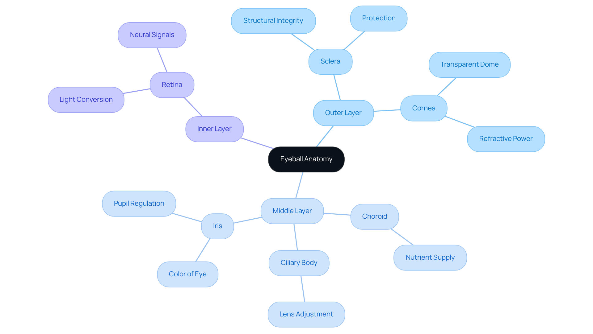
The eyeball anatomy is a remarkable organ essential for sight, which is characterized by its roughly spherical shape and is composed of three primary layers:
- The outer layer, including the sclera and cornea
- The middle layer, consisting of the choroid, ciliary body, and iris
- The inner layer, known as the retina
We understand that this anatomy can feel overwhelming, but each part plays a vital role in your vision.
The sclera, the eye’s white outer covering, provides . The cornea, a transparent dome at the front, allows light to enter while also contributing to the eye’s refractive power. Beneath the sclera lies the choroid, a layer rich in blood vessels that supplies nutrients to the eye, ensuring its health and functionality. The ciliary body, connected to the lens, is crucial for adjusting its shape to help you focus, while the iris, which gives the eye its distinctive color, regulates light entry by controlling the size of the pupil.
Finally, the retina, the innermost layer, is responsible for converting light into neural signals that the brain interprets as images. For cataract patients considering advanced therapies like the Light Adjustable Lens (LAL), understanding this anatomy is essential. It’s common to feel uncertain about what lies ahead, but we are here to support you.
Common symptoms of cataracts include blurred eyesight, difficulty seeing at night, and sensitivity to light. It’s important to recognize how different eye ailments, including cataracts and glaucoma, can impact your vision and overall eye wellness. Resources like the Eye Condition Library can enhance your understanding and education about these conditions. Remember, seeking professional diagnosis for any symptoms you experience is a crucial step towards better eye health.

Understand the Key Components: Cornea, Lens, Retina, and More
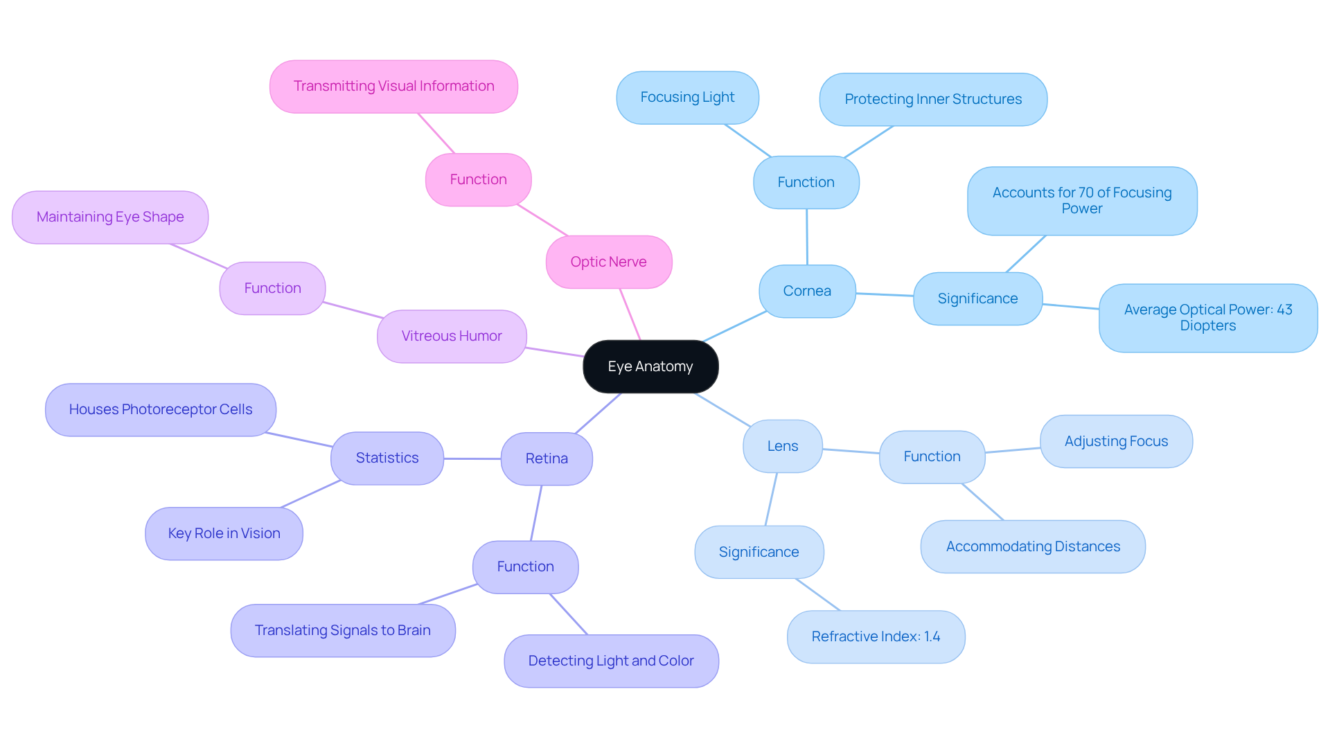
The cornea serves as the eye’s outermost layer, and it’s crucial for focusing light as it enters. We understand that its sensitivity is vital for protecting the inner structures of your eye. Positioned behind the iris, the lens further refines light focus onto the retina. This adaptable structure changes shape to accommodate both near and distant sight, a process essential for clarity. The retina, the innermost layer, houses photoreceptor cells—rods and cones—that detect light and color, translating them into visual signals.
Additionally, the vitreous humor, a gel-like substance, fills the eye and helps maintain its shape, while the optic nerve transmits visual information to the brain. Understanding the components of is vital for recognizing their interactions and the potential implications of dysfunction. It’s common to feel concerned about vision health; for instance, retinal dysfunction can lead to significant vision impairment, affecting millions. Approximately 12 million Americans aged 40 and above experience vision impairment, and about 4.2 million of these individuals are visually impaired. This highlights the significance of regular eye examinations, as they are a proactive measure towards preserving your eye well-being.
Recent studies have shown that the cornea accounts for about 70% of the eye’s total focusing power, with an average optical power of around 43 diopters. The lens plays a critical role in adjusting focus for varying distances, with a refractive index of approximately 1.4. As stated by Northwest Eye, “Understanding these components is crucial, as it helps us recognize how they function together and identify potential vision-related issues.” This knowledge enables you to take proactive measures in preserving your eye well-being.

Link Eye Anatomy to Common Conditions: Cataracts, Glaucoma, and Beyond
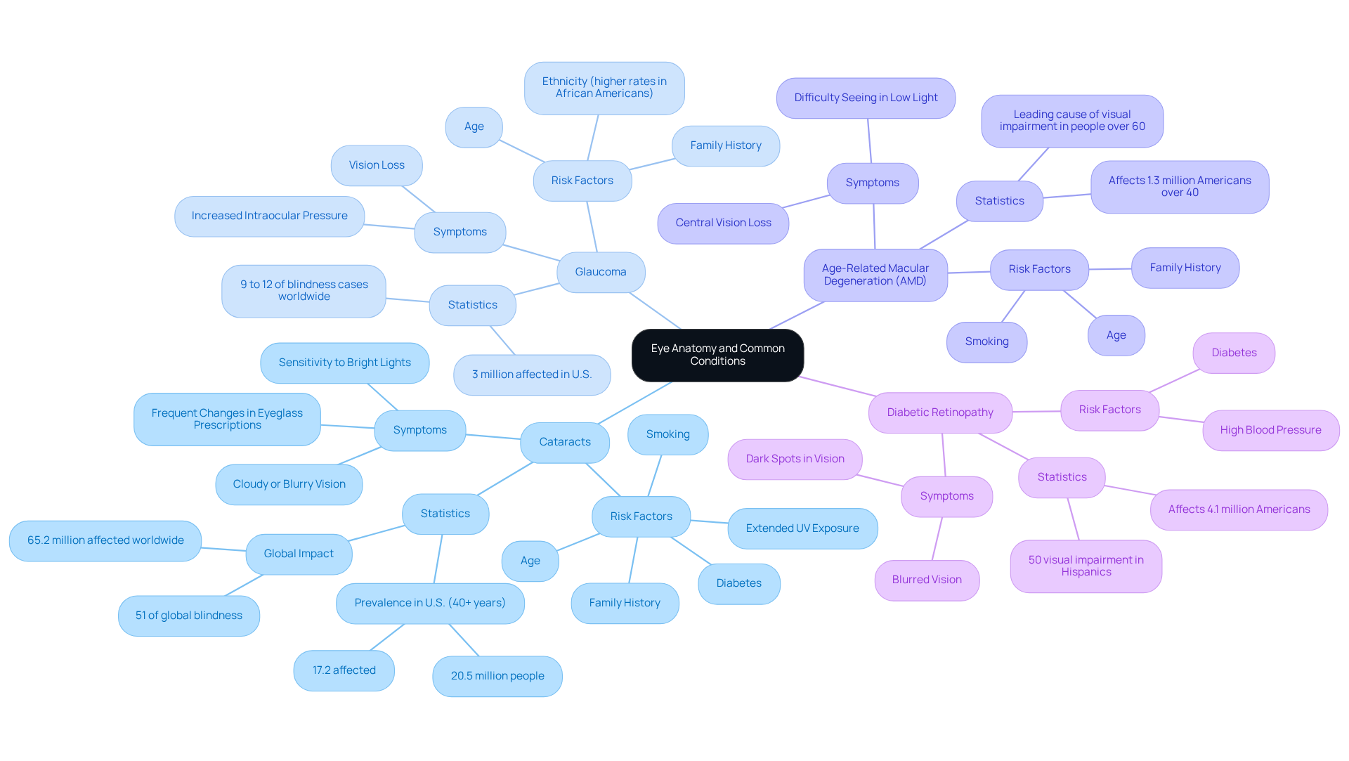
Cataracts occur when the lens of the eye becomes cloudy, leading to unclear sight. This condition is primarily linked to age, affecting about 17.2% of Americans over 40. However, it can also result from factors such as diabetes, extended UV exposure, and specific medical issues. In fact, cataracts account for about 51% of global blindness, impacting around 65.2 million people worldwide. Symptoms may include:
- Cloudy or blurry vision
- Sensitivity to bright lights
- Frequent changes in eyeglass prescriptions
We understand that evaluating your risk for cataracts can be daunting, which is why we encourage you to consider taking our Cataract Self-Test. This tool can assist you in comprehending your eye condition more effectively.
At Northwest Eye, we recognize that accessing eye care services can be a concern for many patients. That’s why we offer a variety of financing programs and payment plans designed to make the cost of care easy and manageable. Our commitment to enhancing eye health is evident in our patient-focused approach, ensuring that everyone has the opportunity to receive the care they need, particularly for issues such as cataracts.
Conversely, glaucoma is characterized by increased intraocular pressure, which can damage the optic nerve and lead to irreversible vision loss. This condition often progresses without apparent symptoms, making crucial for early detection. Around 3 million individuals in the U.S. are affected by glaucoma, many of whom are unaware of their situation. Risk factors include:
- Age
- Family history
- Specific medical conditions, particularly among African Americans, who face elevated rates at younger ages
Other common eye issues include age-related macular degeneration (AMD), which affects the retina and can lead to central vision loss, and diabetic retinopathy, a complication of diabetes that harms retinal blood vessels. Understanding these conditions in relation to eyeball anatomy is crucial, as it empowers individuals to recognize symptoms and seek timely medical intervention. This ultimately preserves their vision and eye health. Furthermore, it’s important to note that 99% of people with cataracts live in underserved communities, highlighting the urgent need for improved access to eye care services.

Conclusion
Understanding the intricate anatomy of the eyeball is essential for grasping how vision functions and the implications of various eye conditions. Each layer of the eye—from the protective sclera to the light-sensitive retina—plays a crucial role in visual perception. We understand that recognizing these components not only enhances knowledge but also empowers you to take proactive steps toward maintaining your eye health.
This article delves into the essential structures of the eyeball, highlighting the significance of the cornea, lens, and retina in focusing light and converting it into visual signals. It’s common to feel overwhelmed by eye health issues, so we also address common conditions such as cataracts and glaucoma, emphasizing the importance of early detection and regular eye examinations. With millions affected by these conditions, understanding their symptoms and risk factors is vital for timely intervention and effective management.
Ultimately, awareness of eyeball anatomy and its link to common eye diseases fosters a deeper appreciation for vision health. We encourage you to seek regular check-ups and utilize available resources to educate yourself about your eye conditions. By doing so, you can ensure better outcomes for your vision and overall well-being, reinforcing the notion that informed patients are empowered patients.
Frequently Asked Questions
What are the main layers of the eyeball anatomy?
The eyeball is composed of three primary layers: the outer layer (sclera and cornea), the middle layer (choroid, ciliary body, and iris), and the inner layer (retina).
What is the function of the sclera?
The sclera, which is the white outer covering of the eye, provides structural integrity and protection for the eyeball.
What role does the cornea play in vision?
The cornea is a transparent dome at the front of the eye that allows light to enter and contributes to the eye’s refractive power.
What is the purpose of the choroid?
The choroid is a layer rich in blood vessels that supplies nutrients to the eye, ensuring its health and functionality.
How does the ciliary body assist in vision?
The ciliary body is connected to the lens and is crucial for adjusting its shape to help focus light on the retina.
What is the function of the iris?
The iris gives the eye its distinctive color and regulates light entry by controlling the size of the pupil.
What does the retina do?
The retina is responsible for converting light into neural signals that the brain interprets as images.
What are common symptoms of cataracts?
Common symptoms of cataracts include blurred eyesight, difficulty seeing at night, and sensitivity to light.
Why is it important to understand eyeball anatomy for cataract patients?
Understanding eyeball anatomy is essential for cataract patients considering advanced therapies like the Light Adjustable Lens (LAL), as it helps them make informed decisions about their treatment.
Where can I find more information about eye conditions?
Resources like the Eye Condition Library can enhance your understanding and education about various eye conditions, including cataracts and glaucoma.


