Posted by: Northwest Eye in General on August 29, 2025
Overview
Central retinal vein occlusion (CRVO) is a serious condition that can significantly impact your vision. We understand that learning about the causes can be concerning. CRVO is primarily associated with risk factors such as:
- Hypertension
- Diabetes
- Atherosclerosis
- Increased intraocular pressure
- Age
These factors contribute to the obstruction of the central retinal vein, which can lead to vision impairment.
It’s common to feel worried about how these conditions and lifestyle choices may affect your health. This article highlights the importance of managing underlying health issues to prevent the occurrence of CRVO. By addressing these risk factors, you can take proactive steps towards protecting your vision and overall well-being. Remember, we are here to help you through this process and support you in making informed decisions about your health.
Introduction
Central retinal vein occlusion (CRVO) is a significant concern in eye health, and we understand how worrying it can be. If not addressed promptly, CRVO can lead to severe vision impairment. This condition arises when the central retinal vein becomes obstructed, often linked to underlying health issues such as hypertension and diabetes, affecting millions worldwide. With the potential for serious complications, it’s crucial to understand the causes, symptoms, and available treatments.
We want you to feel informed and empowered to navigate the complexities of CRVO and safeguard your vision. What should you know to effectively manage this condition?
Define Central Retinal Vein Occlusion
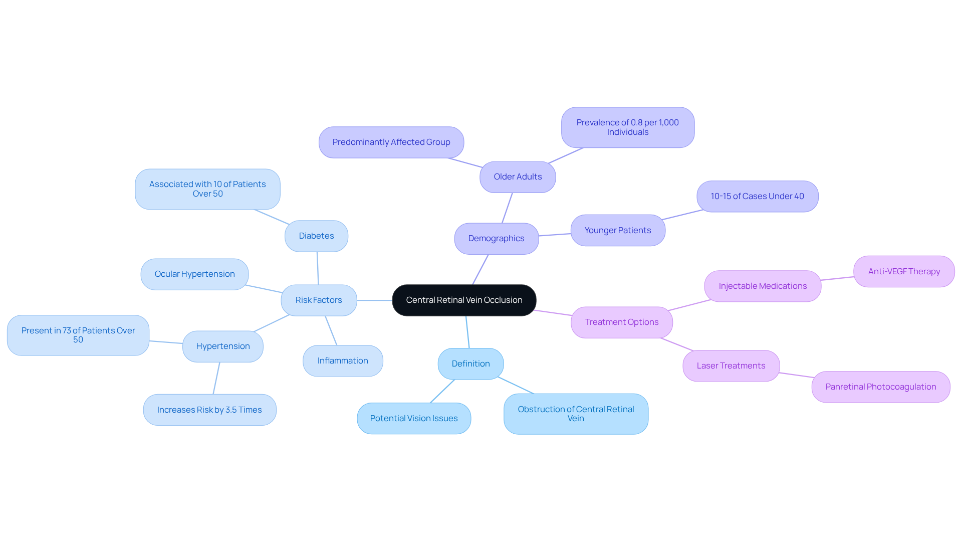
Central retinal vein occlusion is a concerning diagnosis, as it involves an obstruction of the central retinal vein, which is crucial for draining fluid from the retina. This blockage, whether partial or complete, can lead to an accumulation of blood and fluid, potentially causing vision issues. Typically affecting one eye, it is important to address central retinal vein occlusion promptly, since it can lead to significant visual impairment.
We understand that many individuals may feel anxious about this condition, especially since it is often linked to like hypertension and diabetes. These conditions can significantly increase the risk of developing retinal vein occlusion. Recent studies reveal that central retinal vein occlusion is the second most common retinal vascular disorder, affecting an estimated 0.8 per 1,000 individuals, predominantly impacting older adults, particularly those over the age of 50. In fact, about 73% of patients over 50 with this condition also have hypertension, emphasizing the importance of managing blood pressure to reduce risks.
Treatment options for central retinal vein occlusion primarily focus on addressing complications like macular edema. Therapies may include injectable medications and laser treatments like panretinal photocoagulation. We are here to help you through this process, and understanding the demographics and risk factors associated with central retinal vein occlusion is crucial for early detection and effective management. Remember, prompt intervention can significantly improve visual outcomes, and you are not alone in this journey.
Explore Causes of Central Retinal Vein Occlusion
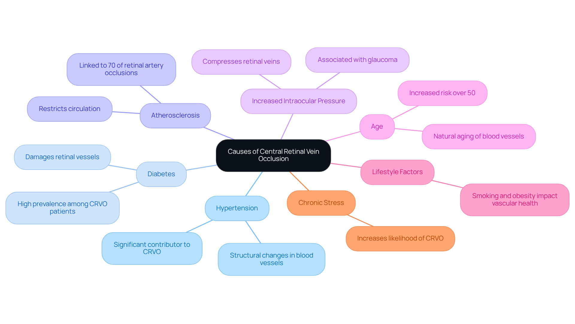
Central retinal vein occlusion is a condition that can profoundly affect eye health, and we recognize how concerning this may be for you. Various risk factors contribute to its occurrence, and being aware of them is crucial for your well-being. Here are some key contributors:
- Hypertension: Elevated blood pressure can cause structural changes in blood vessels, increasing the risk of occlusion. It’s important to know that hypertension is a significant factor for many individuals affected by this condition, with a considerable percentage having a history of elevated pressure.
- Diabetes: This chronic condition can damage retinal vessels, making them more vulnerable to blockages. The prevalence of diabetes among those with central retinal vein occlusion is notably high, highlighting the need to manage sugar levels effectively.
- Atherosclerosis: The hardening of arteries can restrict circulation and lead to clot formation, which is essential in the development of central retinal vein occlusion. Research indicates that up to 70% of retinal artery occlusions are associated with atherosclerosis in the carotid artery, showing its importance.
- Increased Intraocular Pressure: Conditions like glaucoma can raise intraocular pressure, compressing the retinal veins and increasing the likelihood of occlusion.
- Age: The risk of central retinal vein occlusion tends to increase with age, especially in individuals over 50, as a result of the natural aging process of blood vessels.
- Lifestyle Factors: Habits such as smoking and obesity can negatively affect vascular health, further elevating the risk of developing central retinal vein occlusion.
- Chronic Stress: This can also increase the risk of retinal vein occlusion, adding another layer to the complexity of the situation.
Understanding these risk factors is essential for early identification and action. If left untreated, central retinal vein occlusion can result in serious complications, such as loss of sight. We encourage you to prioritize and proactive management of any underlying health issues to preserve your eye health.
It’s important to note that central retinal vein occlusion affected 16.4 million people worldwide in 2008, underscoring the need for awareness and timely intervention. Additionally, statistics show that only 18-41% of eyes improve spontaneously, with visual acuity not improving to 6/12 on average. This reinforces the importance of seeking medical attention promptly. We are here to help you through this process, ensuring you receive the care and support you need.
Identify Symptoms of Central Retinal Vein Occlusion
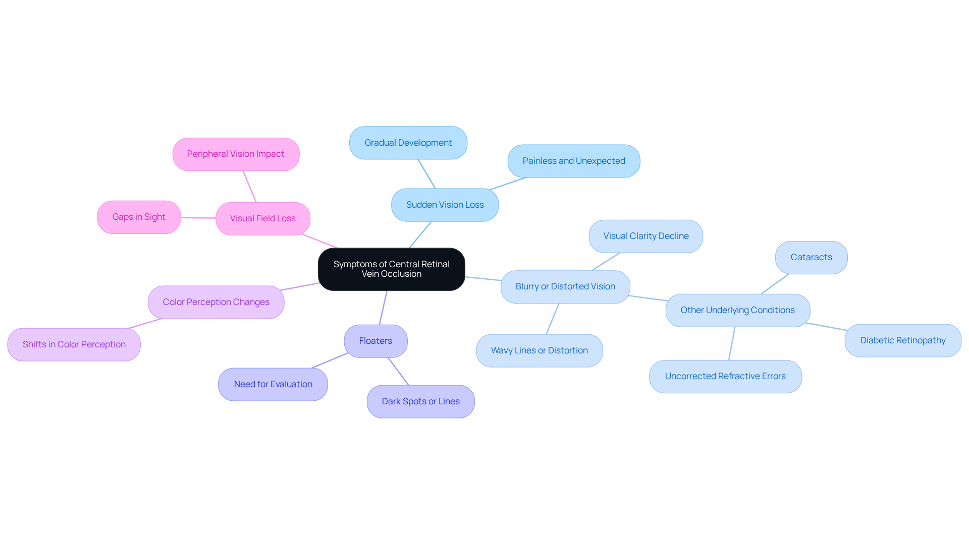
Symptoms of central retinal vein occlusion can manifest in various ways, often leading to significant concerns about vision. We understand how alarming these changes can be, and recognizing the key symptoms is crucial for your peace of mind:
- Sudden Vision Loss: This symptom can appear unexpectedly and is typically painless. It may develop gradually over hours or days, which can be particularly unsettling.
- Blurry or Distorted Vision: Many patients report a decline in visual clarity, often describing their experience as seeing wavy lines or distortion in their visual field. It’s important to note that blurred sight can also stem from other underlying conditions, such as cataracts, diabetic retinopathy, or uncorrected refractive errors, which may intensify the symptoms of central retinal vein occlusion (CRVO).
- Floaters: The sudden appearance of dark spots or lines can indicate changes in the retina, prompting the need for further evaluation.
- Color Perception Changes: Some individuals may notice shifts in how they perceive colors, which can feel disconcerting.
- Visual Field Loss: You might experience noticeable gaps in your sight, particularly affecting your peripheral vision, which can make daily activities more challenging.
Understanding these symptoms is essential, as can lead to prompt medical care, potentially preventing further vision loss. If you are facing any of these symptoms, we encourage you to consult your eye care specialist as soon as possible. Discussing your situation and exploring treatment options can provide reassurance and clarity. Remember, unclear sight can indicate serious underlying issues, and neglecting these symptoms may lead to complications like glaucoma and macular edema. Routine eye examinations are vital for maintaining your eye health and avoiding complications from conditions such as central retinal vein occlusion (CRVO). We are here to help you through this process.
Understand Diagnostic Procedures for CRVO
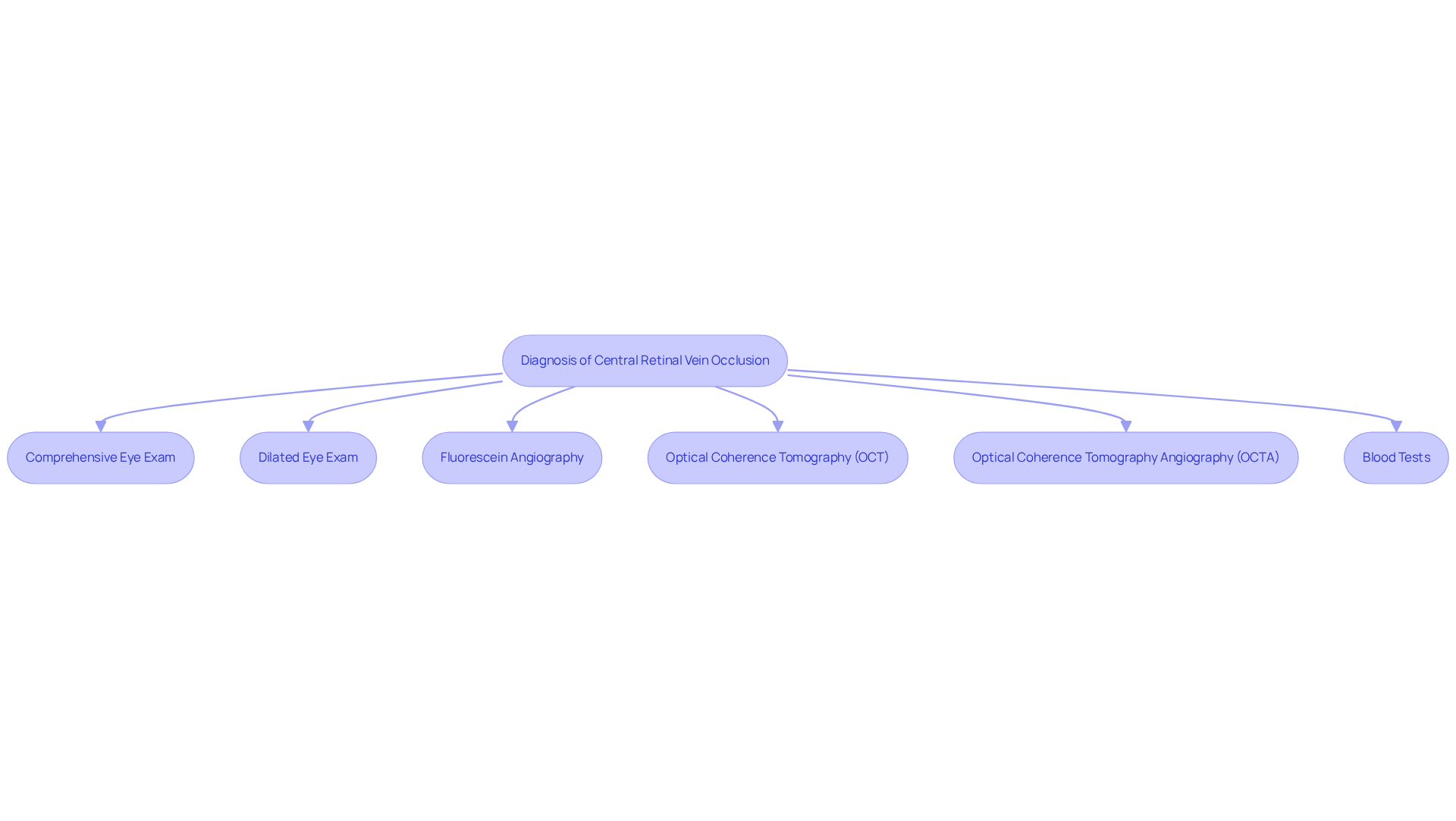
Diagnosing central retinal vein occlusion involves several essential procedures that ensure accurate identification of the condition. We understand that navigating this process can be overwhelming, and we are here to help you through it.
- Comprehensive Eye Exam: An ophthalmologist conducts a thorough examination, including visual acuity tests to assess your vision. This step is vital in understanding your unique situation.
- Dilated Eye Exam: Eye drops are administered to widen your pupils, allowing for a clearer view of the retina and optic nerve. This is crucial for detecting any abnormalities that may be present.
- Fluorescein Angiography: This procedure involves injecting a dye into your bloodstream to visualize blood flow in the retina. It is particularly effective in identifying areas of blockage and assessing the extent of retinal damage. As Dr. Lakshmana M Kooragayala noted, “Fluorescein angiography is the most useful test for the evaluation of retinal capillary nonperfusion and guiding treatment decisions.”
- Optical Coherence Tomography (OCT): This technology provides high-resolution cross-sectional images of the retina, highlighting swelling and other structural changes that may indicate central retinal vein occlusion. This technology allows for a deeper understanding of your retinal health.
- Optical Coherence Tomography Angiography (OCTA): This advanced technique allows for detailed visualization of superficial and deep retinal capillary plexus and choroidal capillary structures without the need for dye injection, enhancing the diagnostic process.
- Blood Tests: Blood tests assist in assessing underlying health issues, such as diabetes or hypertension, which can lead to the onset of central retinal vein occlusion. We know that understanding your overall health is essential in this journey.
Recent advancements in diagnostic techniques have further enhanced the precision of evaluations for central retinal vein occlusion. Research shows that a dilated eye examination is an essential measure in detecting central retinal vein occlusion, since it can reveal signs of blood leakage and other complications. Furthermore, it is crucial to mention that 6-17% of individuals with central retinal vein occlusion may experience blockage of the vein in the other eye, emphasizing the necessity for continuous observation.
Specialists highlight the significance of ; addressing central retinal vein occlusion promptly can greatly lower the chance of sight impairment. This underscores the role of advanced imaging techniques in the comprehensive evaluation of retinal health. Remember, you are not alone in this process, and we are committed to supporting you every step of the way.
Review Treatment Options for Central Retinal Vein Occlusion
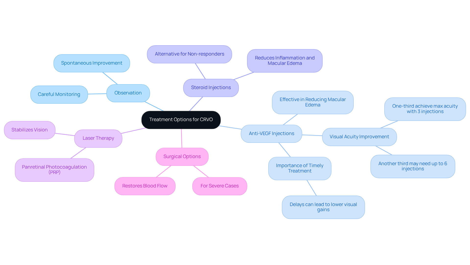
Treatment options for Central Retinal Vein Occlusion (CRVO) are thoughtfully tailored to the severity of the condition, with several approaches available to support you:
- Observation: In mild cases, we understand that careful monitoring can often be sufficient. Many patients may even experience spontaneous improvement in their condition, which can be reassuring.
- Anti-VEGF Injections: Medications like bevacizumab (Avastin) and ranibizumab (Lucentis) are commonly administered via injection into the eye. These treatments have proven to be effective in decreasing macular edema and preventing further loss of sight. Current data shows that over a third of patients achieve maximum visual acuity with just three injections. Another third may need up to six consecutive injections to reach optimal outcomes. Importantly, macular edema (MO) is the most frequent complication from central retinal vein occlusion (CRVO), and anti-VEGF therapy has significantly enhanced vision in eyes affected by macular edema related to central retinal vein occlusion.
- Steroid Injections: For those who may not respond adequately to anti-VEGF therapy, intravitreal steroid injections can effectively reduce inflammation and macular edema related to central retinal vein occlusion, providing an alternative solution.
- Laser Therapy: Panretinal photocoagulation (PRP) is utilized to address complications such as neovascularization that can arise from CRVO. This procedure helps stabilize vision by treating areas of the retina that are not receiving sufficient circulation.
- Surgical Options: In more severe cases, surgical interventions may be necessary to restore blood flow or manage complications effectively. These options are considered when other treatments do not yield satisfactory results.
Recent advancements in the treatment of central retinal vein occlusion, especially in 2025, highlight the ongoing evolution of anti-VEGF therapies. New formulations and delivery methods are continuously enhancing patient outcomes. The efficacy and safety of these treatments for central retinal vein occlusion have been reinforced by extensive clinical trials, establishing anti-VEGF injections as a cornerstone in managing this condition. We emphasize the importance of initiating treatment promptly, as delays of up to six months can lead to compared to immediate intervention. As our understanding of CRVO grows, so does the potential for improved visual outcomes through timely and appropriate interventions. It’s crucial to note that at least 20% of patients may experience poor visual outcomes with severe neovascular complications, underscoring the need for effective treatment strategies. We are here to help you through this process, providing support and guidance every step of the way.
Conclusion
Understanding central retinal vein occlusion (CRVO) is essential for maintaining your eye health and preventing potential vision loss. This condition, characterized by the blockage of the central retinal vein, can lead to serious complications if not addressed promptly. By learning about CRVO’s causes, symptoms, and treatment options, you can take proactive steps in managing your eye health.
We recognize that the risk factors associated with CRVO, such as hypertension, diabetes, and age, can be concerning. It’s crucial to be aware of symptoms like sudden vision loss, blurry vision, and floaters for early detection. The diagnostic procedures outlined, including comprehensive eye exams and advanced imaging techniques, highlight the importance of seeking medical attention at the first sign of trouble. Moreover, various treatment options, ranging from observation to advanced therapies like anti-VEGF injections, provide pathways to potentially restore vision and mitigate the effects of this condition.
Ultimately, awareness and timely intervention are paramount in the fight against central retinal vein occlusion. We encourage you to prioritize routine eye examinations and actively manage any underlying health conditions. By doing so, you can significantly reduce the risk of CRVO, ensuring a better quality of life and preserving your precious vision. Taking these steps not only enhances your personal well-being but also contributes to a broader culture of proactive health management.
Frequently Asked Questions
What is central retinal vein occlusion (CRVO)?
Central retinal vein occlusion is an obstruction of the central retinal vein, which is essential for draining fluid from the retina. This blockage can cause an accumulation of blood and fluid, leading to vision issues, typically affecting one eye.
What are the common risk factors associated with central retinal vein occlusion?
Common risk factors include hypertension, diabetes, atherosclerosis, increased intraocular pressure, age (especially over 50), lifestyle factors like smoking and obesity, and chronic stress.
How prevalent is central retinal vein occlusion?
Central retinal vein occlusion is the second most common retinal vascular disorder, affecting approximately 0.8 per 1,000 individuals, predominantly older adults, particularly those over the age of 50.
What treatments are available for central retinal vein occlusion?
Treatment options primarily focus on addressing complications such as macular edema and may include injectable medications and laser treatments like panretinal photocoagulation.
Why is it important to manage underlying health issues related to CRVO?
Managing underlying health issues, such as hypertension and diabetes, is crucial as these conditions significantly increase the risk of developing central retinal vein occlusion and can lead to serious complications if left untreated.
What are the potential complications of untreated central retinal vein occlusion?
If left untreated, central retinal vein occlusion can result in serious complications, including significant visual impairment or loss of sight.
What percentage of eyes with central retinal vein occlusion improve spontaneously?
Statistics indicate that only 18-41% of eyes improve spontaneously, with visual acuity not improving to 6/12 on average, highlighting the importance of seeking prompt medical attention.
How many people were affected by central retinal vein occlusion worldwide in 2008?
In 2008, central retinal vein occlusion affected approximately 16.4 million people worldwide, underscoring the need for awareness and timely intervention.


