Posted by: Northwest Eye in General on August 27, 2025
Overview
Choroidal melanoma is a serious eye tumor that develops from melanocytes in the choroid. As the most common primary intraocular cancer in adults, it is crucial to detect it early for better outcomes. We understand that receiving such news can be overwhelming, and we want to assure you that there are steps we can take together.
Accurate diagnosis is essential, and advanced imaging techniques play a vital role in this process. It’s common to feel anxious about what comes next, but knowing your options can provide some peace of mind. Treatment may involve:
- Radiation therapy
- Surgery
tailored specifically to your individual circumstances. We are here to help you through this process and ensure you receive the care you need.
Timely medical intervention is critical in improving survival rates. We encourage you to reach out to healthcare professionals who can guide you in making informed decisions. Remember, you are not alone in this journey; support is available every step of the way.
Introduction
Choroidal melanoma is the most common primary intraocular cancer in adults, and we understand that facing such a diagnosis can be overwhelming. This condition presents a significant health challenge, especially if not detected early, and nearly half of those affected face the risk of metastasis. Therefore, understanding the nuances of diagnosis, treatment options, and long-term care is crucial for improving patient outcomes.
Navigating this complex landscape of ocular health can feel daunting. It’s common to have questions and concerns about what lies ahead. What steps can be taken to ensure timely intervention and optimal care? We are here to help you through this process, offering support and guidance every step of the way.
Define Choroidal Melanoma: Overview and Importance
Choroidal melanoma is a type of malignant growth that arises from the melanocytes in the choroid, the layer of blood vessels and connective tissue located between the retina and the sclera of the eye. As the most prevalent primary intraocular cancer in adults, choroidal melanoma accounts for around 85% of all ocular tumors. We understand that receiving this information can be overwhelming, but early detection is critical. The tumor can often remain asymptomatic for extended periods, leading to late-stage diagnosis and poorer outcomes.
Statistics reveal that nearly half of individuals with uveal cancer will develop metastatic illness, primarily affecting the liver. This highlights the urgent need for prompt intervention. For instance, a study indicated that the median survival for individuals with metastasis is only 3.1 months, emphasizing the importance of early diagnosis. Additionally, the occurrence rates of eye cancer vary globally, with reports showing figures as high as 10 per million in the United Kingdom and 7.6 per million in Australia.
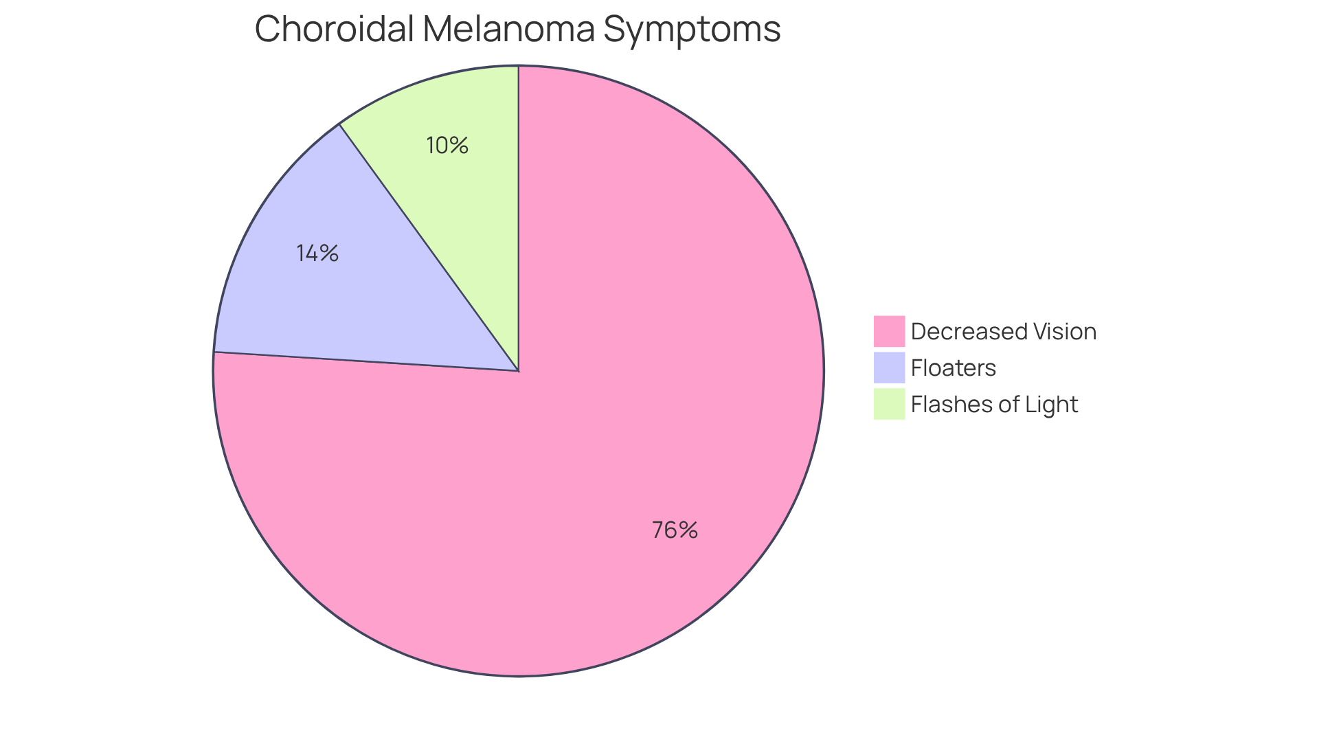
Oncologists emphasize that recognizing symptoms early—such as decreased vision (38%), floaters (7%), or flashes of light—can lead to prompt medical evaluation and treatment, ultimately improving survival rates. It’s common to feel anxious about these symptoms, but grasping the realities of ocular cancer is crucial for both patients and healthcare professionals. This understanding promotes awareness and supports . We are here to help you through this process, ensuring that you feel supported every step of the way.

Diagnose Choroidal Melanoma: Methods and Criteria
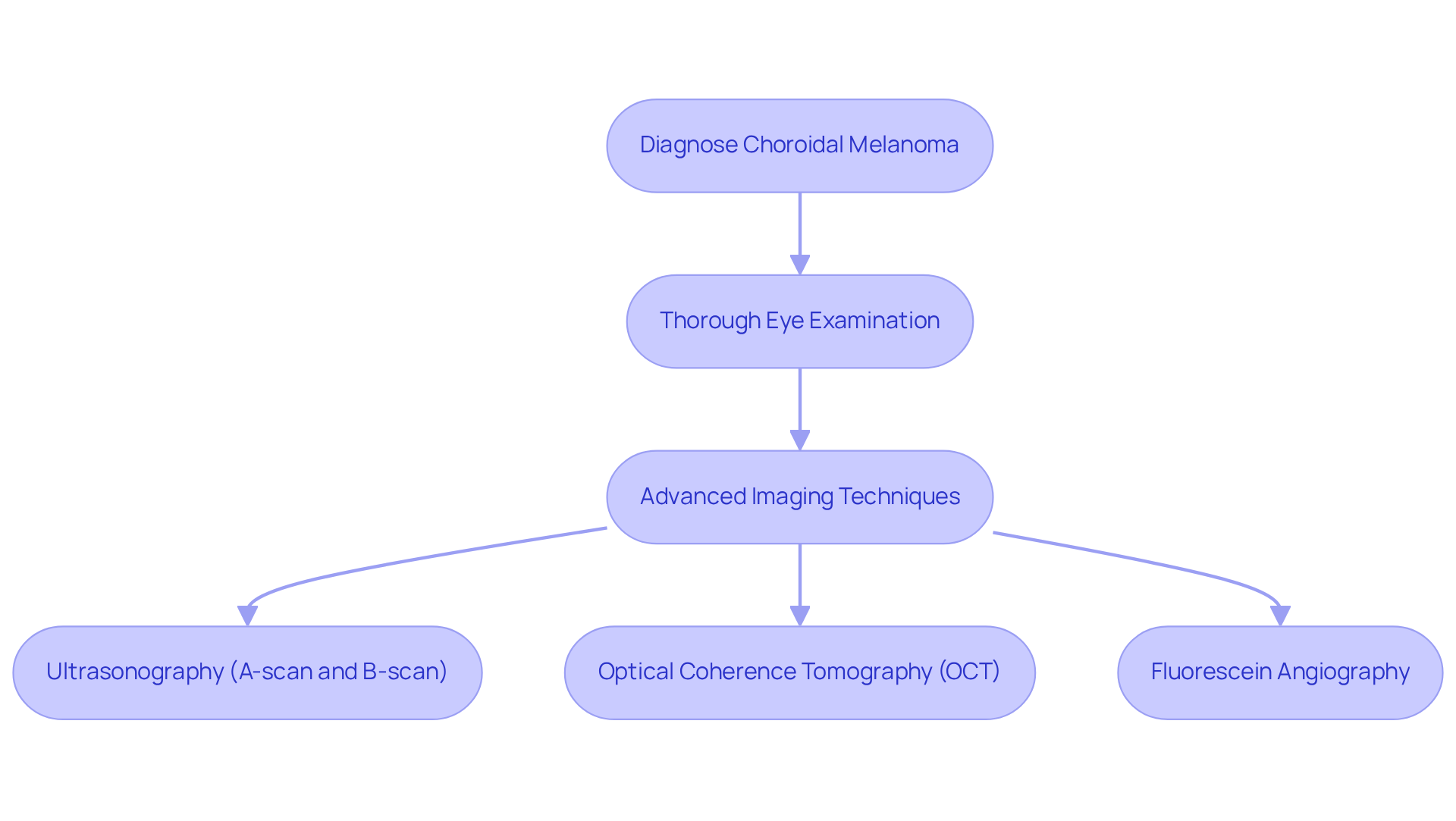
Diagnosing choroidal melanoma can feel overwhelming, but we are here to assist you throughout this process. A thorough eye examination is essential, which includes:
- A dilated fundus examination
- Advanced imaging techniques like:
- Ultrasonography (both A-scan and B-scan)
- Optical coherence tomography (OCT)
- Fluorescein angiography
These imaging techniques are crucial for evaluating key diagnostic factors, such as the mass’s size, shape, and internal reflectivity.
For instance, enhanced depth imaging OCT can visualize tumors smaller than 3 mm, providing critical insights into their characteristics. It’s reassuring to know that the Collaborative Ocular Melanoma Study (COMS) has demonstrated a 99.6% accuracy in diagnosing medium and large-sized choroidal melanoma, underscoring the effectiveness of these imaging techniques.
Frequent eye check-ups are vital, especially for individuals at increased risk, like those with a history of nevi. Early identification significantly enhances treatment outcomes. We understand that this can be a stressful time, but the integration of these diagnostic methods not only aids in the precise characterization of the growths but also informs the selection of . Ultimately, this enhances patient care and provides you with the best possible support.

Treat Choroidal Melanoma: Management Strategies and Options
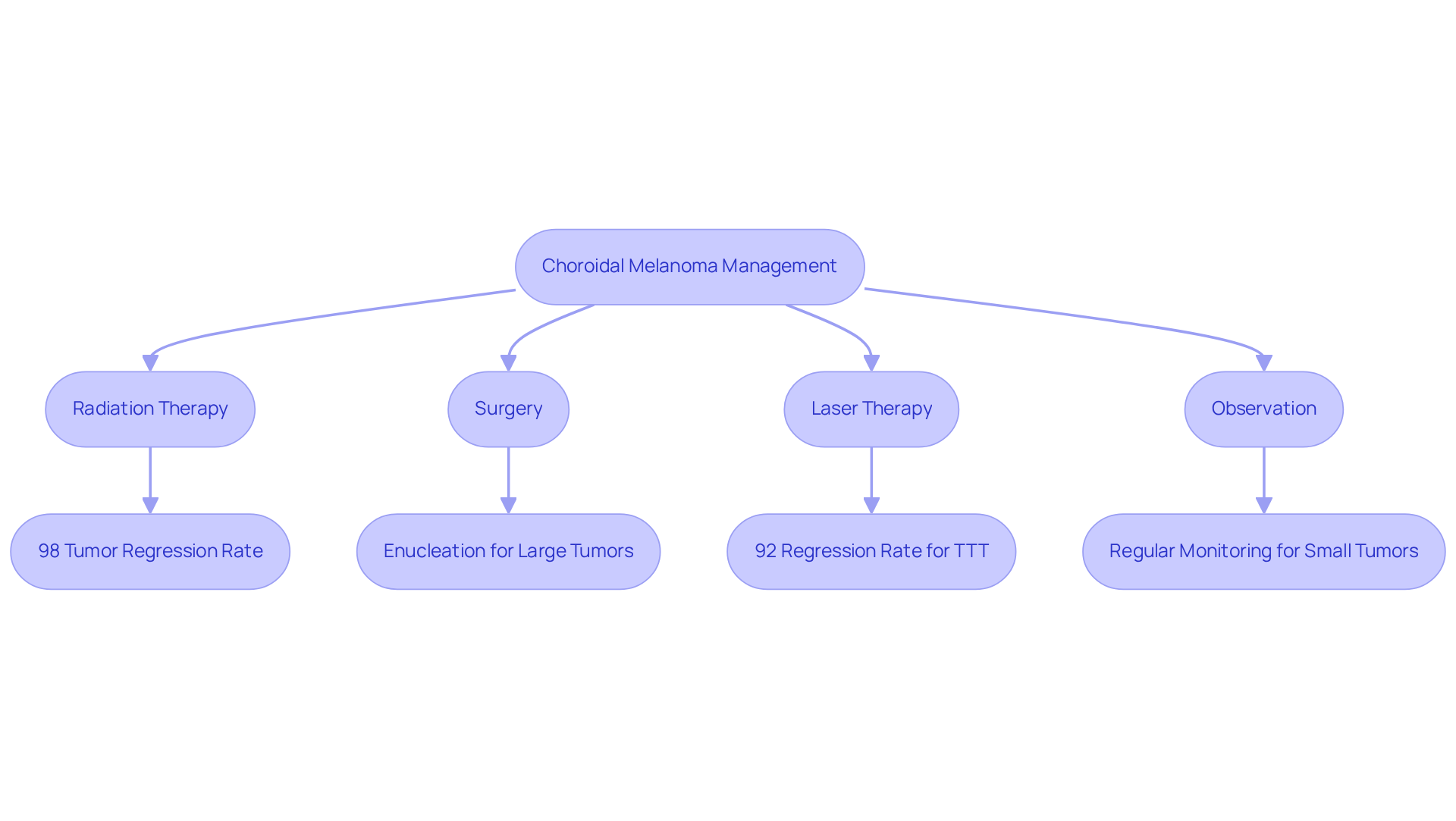
Management approaches for eye cancer are tailored to each individual’s unique circumstances, including the size, location, and presence of metastasis. We understand that receiving a diagnosis can be overwhelming, and we are here to guide you through your options with compassion and care.
- Radiation Therapy: Often the primary treatment for small to medium-sized growths, radiation therapy employs techniques like plaque brachytherapy. This method has an impressive tumor regression rate of 98% for small choroidal melanoma, allowing us to target tumors precisely while preserving surrounding healthy tissue. Research indicates that individuals receiving radiotherapy alone experience a 45% decrease in all-cause mortality compared to those undergoing surgical procedures. The 5-year overall survival rate for patients undergoing radiotherapy is approximately 77.69%, with a 10-year rate of 62.03%. These statistics highlight the effectiveness of this treatment option and the hope it offers.
- Surgery: In cases where growths are larger or causing significant ocular damage, surgical interventions such as local resection or enucleation may be necessary. Enucleation, or the removal of the eye, is typically reserved for situations where other therapies have proven ineffective or when the growth is too large for eye-sparing methods. We know this can be a difficult decision, and we are here to support you every step of the way.
- Laser Therapy: Transpupillary thermotherapy (TTT) is a minimally invasive option that utilizes laser energy to eliminate cancerous cells. While TTT has shown a of 92% in small ocular lesions, it is important to note that it may have elevated local recurrence rates, making it less suitable for larger growths.
- Observation: For small, asymptomatic growths, a watchful waiting approach may be adopted, involving regular monitoring to assess any changes. This strategy is particularly relevant for small eye tumors, where initial observation is often recommended due to diagnostic uncertainty. We understand that waiting can be challenging, but rest assured, we will be here to monitor your progress closely.
Every treatment option has its own advantages and drawbacks, and the selection of management should be personalized according to your specific situation and tumor characteristics. Recent advancements in targeted therapies and ongoing clinical trials continue to shape the field of eye cancer management, providing hope for improved outcomes. Remember, you are not alone in this journey; we are here to help you through this process.

Follow Up and Prognosis: Long-Term Care for Choroidal Melanoma Patients
Long-term follow-up care is essential for individuals treated for eye tumors, as it significantly influences both outcomes and quality of life. We understand that navigating this journey can be challenging, which is why regular check-ups are so important. These typically include:
- Ocular Examinations: We recommend comprehensive eye exams every 3 to 6 months for the first few years following treatment, transitioning to annual exams thereafter. This schedule allows for early detection of any changes in vision or signs of recurrence, providing peace of mind.
- Imaging Studies: Periodic imaging, such as ultrasound or optical coherence tomography (OCT), plays a crucial role in monitoring potential recurrence or metastasis. These tests help visualize any abnormalities that may arise post-treatment, ensuring that nothing goes unnoticed.
- Systemic Monitoring: Given the risk of metastasis, liver function tests and imaging studies, including CT scans, are often recommended to assess for any spread of the disease. It’s important to note that hepatic metastasis occurs in about 50% of uveal cancer patients, making careful monitoring essential for your health.
The outlook for eye cancer can be influenced by various factors, such as mass size and the presence of metastasis at diagnosis. We want to reassure you that and prompt treatment can lead to significantly improved survival rates, with five-year survival rates exceeding 90% for small tumors. Recent studies indicate that the cumulative disease-specific survival for individuals treated for choroidal melanoma remains favorable, especially when managed with a personalized approach.
We are here to help you through this process, and continuous education and support from healthcare providers are paramount in the long-term management of these patients. This ensures that you remain informed and proactive in your care, empowering you to navigate this journey with confidence.

Conclusion
Choroidal melanoma is a significant challenge in ocular health, being the most common primary intraocular cancer in adults. We understand that facing this diagnosis can be overwhelming. Recognizing its implications, from early detection to tailored treatment strategies, is crucial for improving patient outcomes. The emphasis on proactive diagnosis and comprehensive care is paramount, as timely intervention can drastically alter the prognosis for those affected by this serious condition.
Key insights from our discussion highlight the critical nature of recognizing symptoms early, employing advanced diagnostic techniques, and utilizing a variety of treatment options tailored to individual circumstances. It’s common to feel uncertain when confronted with statistics revealing the stark realities of metastasis and survival rates. However, awareness and education play vital roles in navigating the complexities of choroidal melanoma. Continuous follow-up and monitoring are essential components of long-term care, ensuring that patients remain vigilant and supported throughout their journey.
Ultimately, the significance of understanding choroidal melanoma cannot be overstated. By fostering awareness and encouraging proactive health measures, individuals can empower themselves and their healthcare providers to combat this formidable disease effectively. A commitment to ongoing education, timely diagnosis, and personalized care strategies will not only enhance survival rates but also improve the quality of life for those affected by choroidal melanoma. We are here to help you through this process.
Frequently Asked Questions
What is choroidal melanoma?
Choroidal melanoma is a malignant growth that originates from melanocytes in the choroid, which is the layer of blood vessels and connective tissue located between the retina and the sclera of the eye.
How common is choroidal melanoma?
Choroidal melanoma is the most prevalent primary intraocular cancer in adults, accounting for about 85% of all ocular tumors.
Why is early detection of choroidal melanoma important?
Early detection is critical because the tumor can often remain asymptomatic for long periods, leading to late-stage diagnosis and poorer outcomes. Nearly half of individuals with uveal cancer may develop metastatic illness, primarily affecting the liver.
What are the survival rates for individuals with metastatic choroidal melanoma?
The median survival for individuals with metastasis is only 3.1 months, highlighting the urgency of early diagnosis and intervention.
What are the occurrence rates of choroidal melanoma in different regions?
The occurrence rates of eye cancer vary globally, with reports indicating figures as high as 10 per million in the United Kingdom and 7.6 per million in Australia.
What symptoms should individuals be aware of regarding choroidal melanoma?
Early symptoms to recognize include decreased vision (38% of cases), floaters (7%), and flashes of light. Recognizing these symptoms can lead to prompt medical evaluation and treatment.
How can understanding choroidal melanoma help patients and healthcare professionals?
Understanding the realities of ocular cancer promotes awareness, supports proactive health initiatives, and helps in alleviating anxiety for both patients and healthcare professionals.


