





Posted by: Northwest Eye in Eye Symptoms and Their Causes on May 26, 2026
Introduction
Many people find it challenging to understand the complexities of eye health, especially when faced with conditions like the eye bubble. We want to help you understand the causes, symptoms, and diagnostic approaches related to eye bubbles. This condition can significantly impact your vision and recovery after surgery. It’s common to feel concerned about blurred vision and discomfort, so let’s explore how you can recognize the signs of an eye bubble and navigate the path to effective treatment.
Define the Eye Bubble: Importance in Eye Health
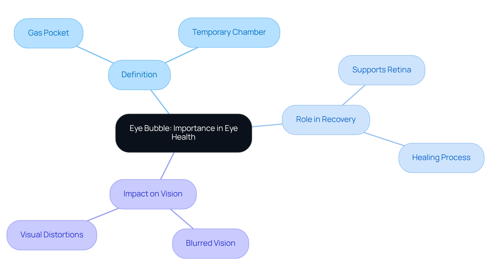
We understand that undergoing eye surgery can be a daunting experience, and it’s natural to have questions about what to expect. An eye cavity, often known as a gas pocket or intraocular space, is a temporary chamber of gas injected into the eye during specific surgical procedures, such as retinal detachment repair. This sac plays a vital role in supporting your retina during recovery, helping to keep it in place as it heals.
Understanding the eye chamber is important because it can affect how clearly you see and how smoothly your recovery goes. It’s common to experience blurred vision or visual distortions while the gas is in place, and these changes can last from several days to weeks, depending on the type of gas used.
Recognizing the importance of this aspect of eye health can help you feel more informed and prepared for your recovery, as it directly impacts your healing and visual outcomes. Remember, we are here to help you through this process.
Explore Causes and Symptoms of the Eye Bubble
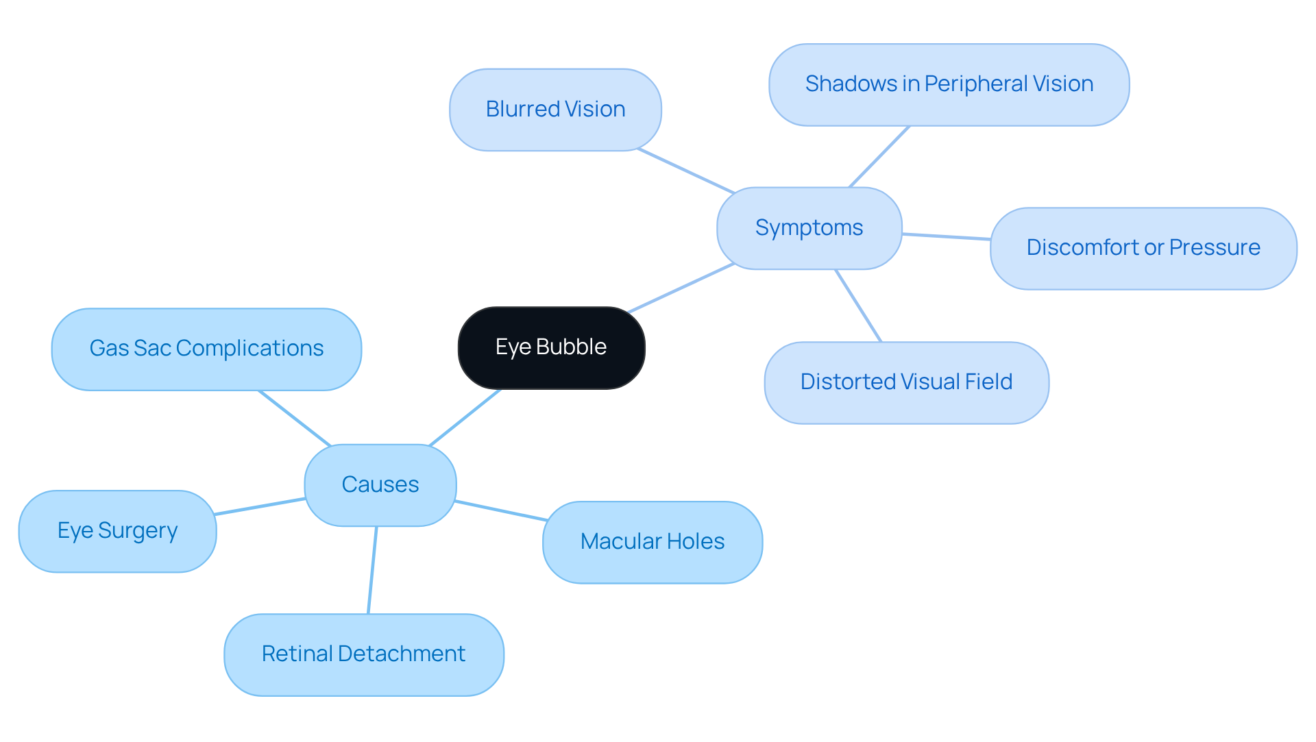
We understand that experiencing an eye bubble can be unsettling and may raise many questions. Eye distortions, including the occurrence of an eye bubble, can arise from several conditions, such as retinal detachment, macular holes, and certain types of eye surgery. You might notice symptoms like blurred vision, a distorted visual field, or even shadows in your peripheral vision. In some cases, discomfort or a sensation of pressure in the eye may accompany these symptoms.
Blurred vision can stem from several issues. These include nearsightedness, farsightedness, presbyopia, and astigmatism. More serious conditions like cataracts, diabetic retinopathy, dry eyes, and macular degeneration can also be culprits. It can be overwhelming to identify these symptoms on your own, but recognizing them is important, as they can indicate underlying issues that require prompt medical evaluation.
For example, a gas sac used during retinal surgery is designed to support the retina. However, if it lasts longer than anticipated or is accompanied by intense pain, it may indicate complications that require attention from an eye care specialist. If you notice blurred vision or any other concerning symptoms, please reach out for professional medical help as soon as you can. By taking action now, you can safeguard your vision and promote better eye health.
Detail Diagnostic Approaches for Eye Bubble Assessment
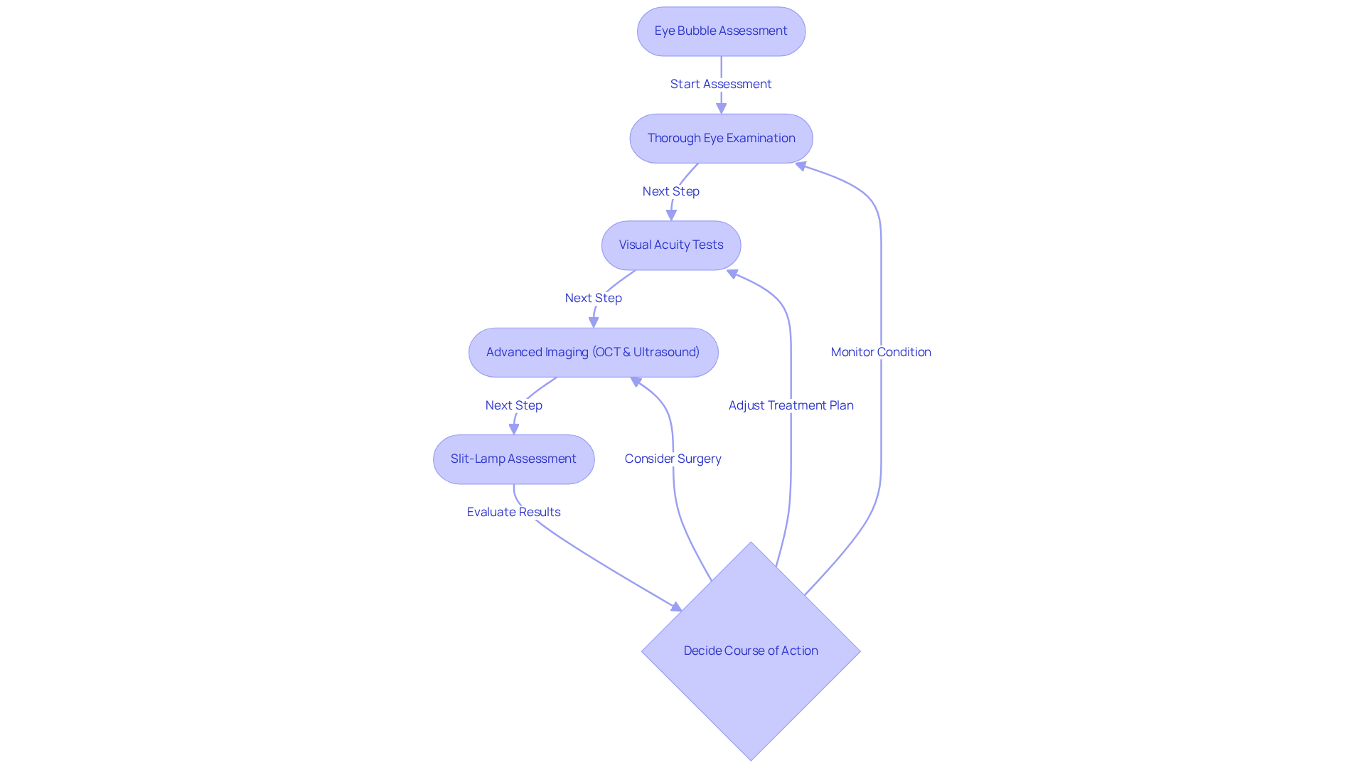
We understand that dealing with eye bubble conditions can be overwhelming, and knowing how to identify potential problems is crucial for your health. Identifying issues with your eyes can be challenging, and a thorough eye examination, including visual acuity tests and advanced imaging like OCT and ultrasound, is essential for understanding your eye bubble. These technologies help us assess the structure and function of your eyes, making it easier to spot potential complications, including conditions like an eye bubble or retinal detachment. OCT is especially effective for diagnosing different eye conditions, including issues related to eye bubbles, giving us high-resolution images that help us detect problems early and plan your treatment accordingly. As one leading ophthalmologist puts it, ‘OCT is invaluable in our diagnostic toolkit, enabling us to catch conditions early and tailor treatment effectively.’
During your examination, we may also perform a slit-lamp assessment to check for any abnormalities related to the eye bubble in the front part of your eye. Using these diagnostic methods is crucial for deciding the best course of action regarding the eye bubble. This might mean monitoring your condition, adjusting your treatment plan, or considering surgery if complications arise.
It’s concerning to know that nearly 900,000 Americans are at risk for vision-threatening diabetic retinopathy, and many more will face cataracts in the coming years. Accurate imaging is vital for maintaining your eye bubble health, as it significantly impacts your overall well-being and quality of life.
Review Treatment Options for Eye Bubble Management
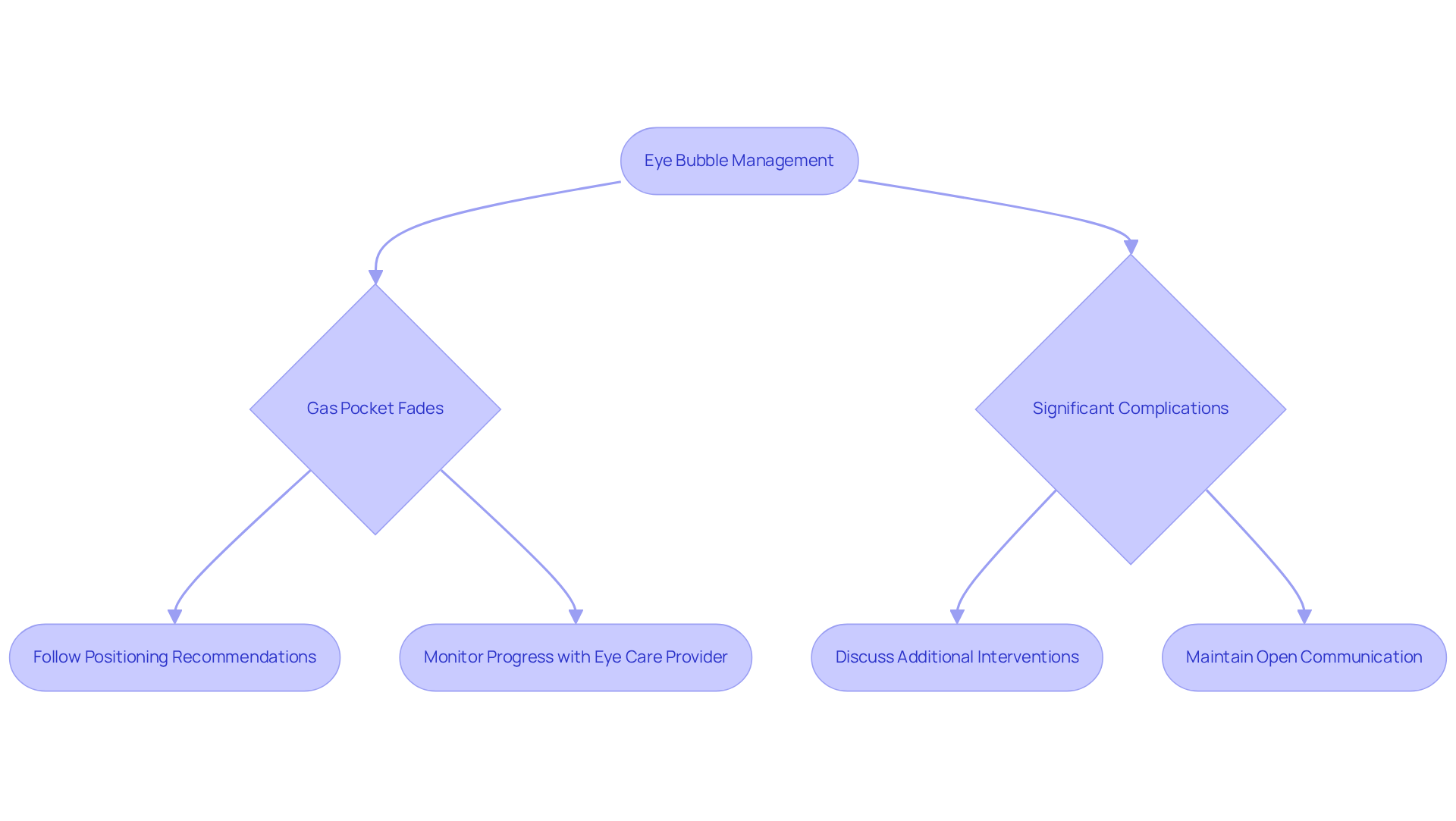
We understand that facing eye issues can be overwhelming, and it’s important to explore all your treatment options. Treatment alternatives for addressing eye issues depend on the underlying cause and your specific circumstances. In many cases, the gas pocket may gradually fade, and we encourage you to follow specific positioning recommendations to support your recovery.
For instance, after retinal surgery, you might need to maintain a face-down position to help the gas effectively support the retina. We know that dealing with visual disturbances can be distressing. If the gas pocket causes significant complications, additional interventions may be necessary. This may lead to further discussions with your eye care provider about the best options for your situation.
It’s vital to maintain an open line of communication with your eye care provider to monitor the eye bubble’s progress and discuss any concerns that may arise during your recovery. By staying connected with your eye care provider, you can navigate your recovery with confidence and support.
Conclusion
We understand that navigating the world of eye surgery and visual disturbances can be daunting, but knowing about the eye bubble can help ease your concerns. This temporary gas pocket is here to support your retina during recovery, and we want you to know how it can influence your healing journey. By understanding what an eye bubble is and how it affects your recovery, you can feel more in control of your eye health journey.
The article explored the various aspects of eye bubbles, including:
- Causes
- Symptoms
- Diagnostic methods
- Treatment options
It highlighted that conditions such as retinal detachment and surgical procedures can lead to the formation of an eye bubble, often accompanied by blurred vision and other visual distortions. Diagnostic techniques like OCT and ultrasound are crucial for assessing the health of the eye bubble and planning appropriate interventions. Treatment may involve monitoring the gas pocket or adhering to specific positioning guidelines to ensure proper healing.
Taking proactive steps can not only ease your worries but also pave the way for a smoother recovery process. Engaging with eye care professionals and understanding the signs and symptoms associated with eye bubbles can empower you to take charge of your eye health. By taking these steps, you’re not just protecting your vision; you’re also empowering yourself to face this journey with confidence and support.
Frequently Asked Questions
What is the eye bubble?
The eye bubble, also known as an intraocular gas pocket, is a temporary chamber of gas injected into the eye during specific surgical procedures, such as retinal detachment repair. It supports the retina during recovery.
Why is the eye bubble important for eye health?
The eye bubble is crucial because it helps keep the retina in place as it heals, which can significantly impact the healing process and visual outcomes.
What should I expect in terms of vision while the gas bubble is in place?
It is common to experience blurred vision or visual distortions while the gas is in place, and these changes can last from several days to weeks, depending on the type of gas used.
How does understanding the eye bubble help during recovery?
Recognizing the importance of the eye bubble can help patients feel more informed and prepared for their recovery, as it directly affects their healing and visual outcomes.
What types of surgeries typically involve the use of an eye bubble?
The eye bubble is often used in surgical procedures like retinal detachment repair.
List of Sources
- Detail Diagnostic Approaches for Eye Bubble Assessment
- Ophthalmic Case Studies | Ophthalmology and Visual Sciences | Medical College of Wisconsin (https://mcw.edu/departments/ophthalmology-eye-institute/education/ophthalmic-case-studies)
- The Statistics on Eye Disease in America (2022) | NVISION (https://nvisioncenters.com/education/eye-disease-statistics)