





Posted by: Northwest Eye in Retinal Health and Diseases on July 30, 2026
Introduction
We understand that recognizing the symptoms of a horseshoe tear can feel overwhelming, but you’re not alone in this journey. This type of retinal injury, which appears as a U-shaped rupture, can be concerning if not treated promptly. Ignoring these symptoms could lead to serious complications, including vision loss. By exploring the symptoms, treatment options, and follow-up care together, we can help you feel more informed and empowered. What steps can you take to ensure timely intervention and protect your vision?
Define Horseshoe Tear: Understanding the Condition
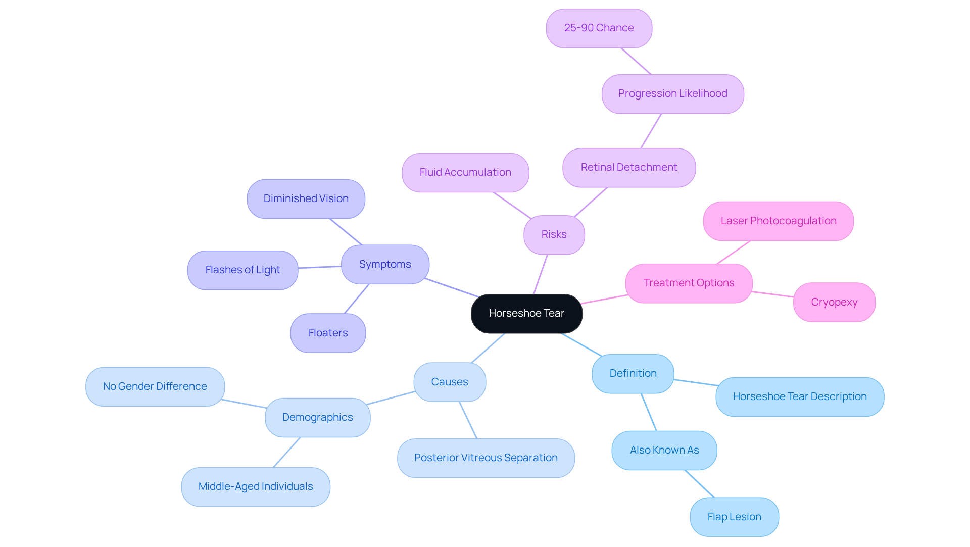
We understand that discovering a horseshoe tear can be concerning, but knowing more about it can help you feel more in control. A horseshoe tear, also referred to as a flap lesion, is a specific kind of retinal injury that appears U-shaped. This condition occurs when the vitreous gel within the eye pulls on the peripheral retina, often due to posterior vitreous separation (PVS). Horseshoe tear lesions are breaks in the neurosensory retina that typically occur at the back of the retina, especially in the superotemporal and superonasal areas. We know that if left untreated, these injuries can lead to serious complications, including retinal separation, which can pose a significant risk to your vision.
It’s common to feel worried about the symptoms associated with this condition. The most frequent reason for a crescent-shaped rupture is a posterior vitreous separation, which can occur in middle-aged individuals, with no significant difference between men and women. Symptoms like floaters, flashes of light, and diminished vision may suggest the presence of a retinal rupture. Timely assessment is essential, as symptomatic crescent-shaped disruptions have a 25 to 90 percent likelihood of advancing to detachment of the inner eye layer if not managed swiftly. It’s important to know that if you’re experiencing symptoms during a posterior vitreous separation, there’s a 15 percent chance of developing an eye injury. Delays in diagnosis can lead to fluid accumulation under the retina, worsening the condition. Therefore, if you have risk factors like high myopia or a history of lattice degeneration, we encourage you to have regular dilated eye examinations, even if you don’t notice any symptoms.
Case studies from the Midwest highlight the importance of timely intervention. For example, one patient exhibited a crescent-shaped rupture along with vitreous floaters, emphasizing the need for thorough management strategies. Another case demonstrated that untreated retinal breaks significantly increase the risk of retinal detachment, underscoring the necessity for prompt treatment.
Taking proactive steps now can make all the difference in preserving your vision and peace of mind. Early detection and treatment, such as laser photocoagulation or cryopexy, can effectively seal the rupture and prevent further complications, ensuring better outcomes for patients.
Identify Symptoms: Recognizing Horseshoe Tear Indicators
We understand that experiencing changes in your vision can be alarming and concerning. Common symptoms of a horseshoe tear include:
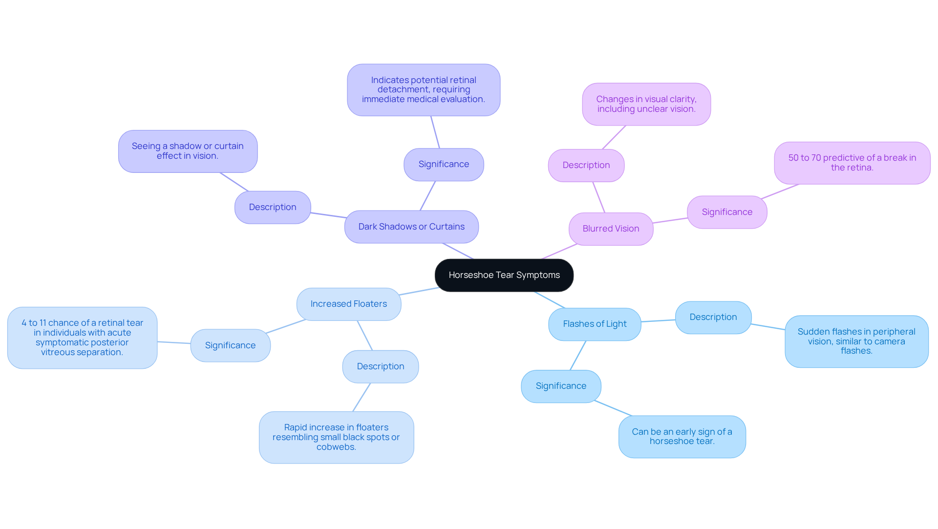
- Flashes of Light: You might notice sudden flashes in your peripheral vision, similar to camera flashes. This can often be an early sign of a horseshoe tear.
- Increased Floaters: A rapid increase in floaters, which may look like small black spots or cobweb-like structures, is a significant warning sign. Research indicates that individuals with acute symptomatic posterior vitreous separation have a 4% to 11% chance of a tear in the eye, making this symptom particularly concerning.
- Dark Shadows or Curtains: Some people report seeing a shadow or curtain effect in their vision, which can indicate a more serious condition, such as retinal detachment. This symptom requires immediate medical evaluation.
- Blurred Vision: Changes in visual clarity, including unclear vision, can also signal a specific type of retinal issue. The presence of vitreous hemorrhage is 50% to 70% predictive of a break in the retina, underscoring the importance of timely assessment.
We want to emphasize how important it is to recognize these symptoms early so you can receive the care you need. It’s important to know that untreated symptoms can lead to serious issues, with retinal detachment occurring in 30% to 50% of cases. Therefore, if you experience any of these symptoms, please seek prompt evaluation from a retina specialist to mitigate the risk of permanent vision loss. As mentioned by RetinaCare, “When crescent-shaped ruptures are identified and addressed swiftly, the result is typically very positive.” Taking action now can make all the difference in preserving your vision for the future.
Explore Treatments: Options for Managing Horseshoe Tears
We know that facing a diagnosis of horseshoe tear can be overwhelming, but understanding your treatment options can bring you peace of mind. Here are some key options to consider:
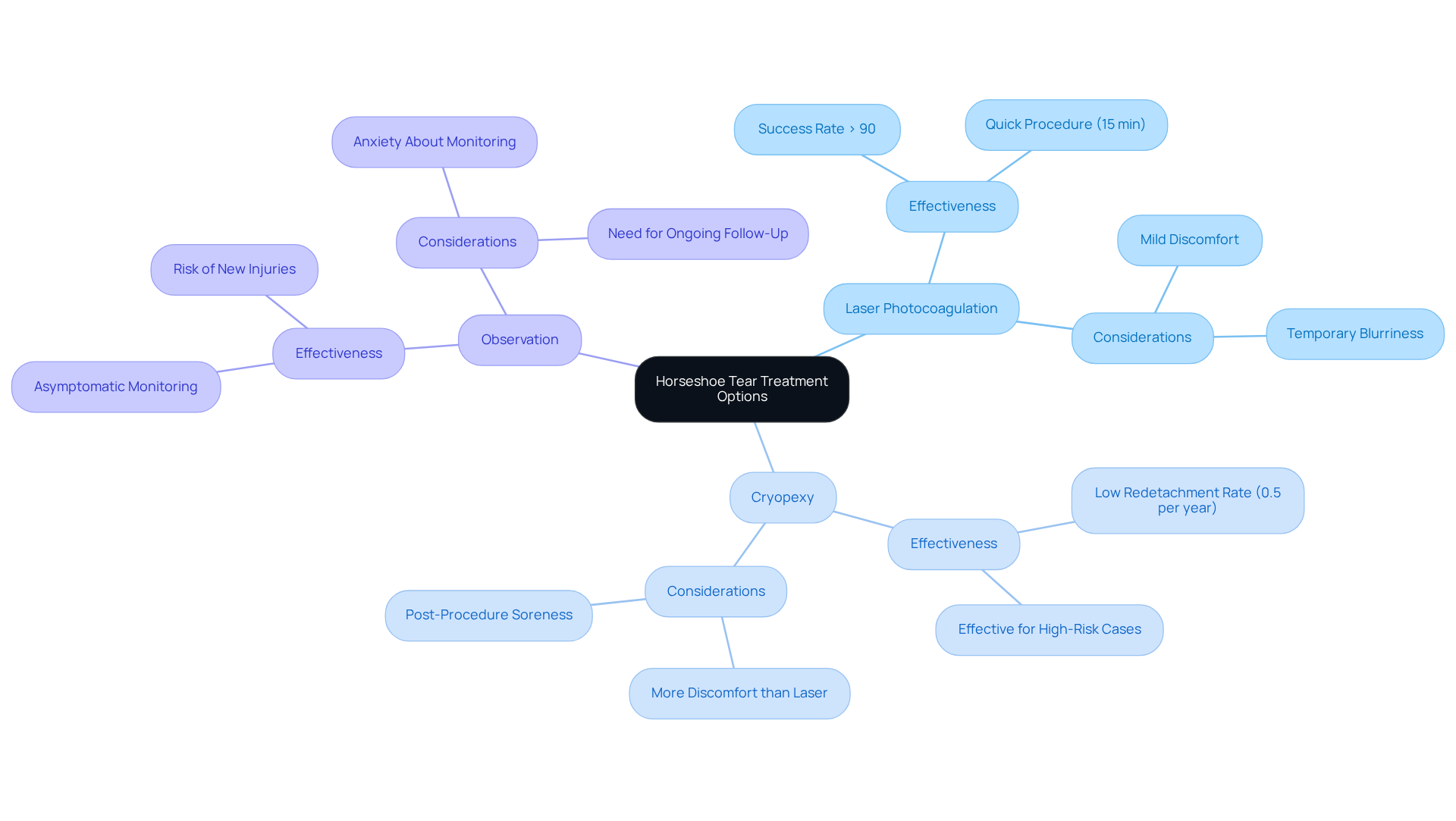
- Laser Photocoagulation: This procedure uses a focused laser beam to create small burns around the tear. This promotes adhesion of the retina to the underlying tissue and helps prevent fluid from entering the tear. With a success rate exceeding 90%, laser photocoagulation is recognized for its effectiveness in sealing eye injuries and preventing complications. Dr. Grace Zhang emphasizes that both laser photocoagulation and cryopexy are safe and effective methods for addressing retinal issues.
- Cryopexy: This technique involves applying extreme cold to the area surrounding the rupture, which helps seal it and prevent further complications. Cryopexy has a low redetachment rate of only 0.5% per year after six months of successful attachment, making it a reliable option for managing retinal issues. In one study, proliferative vitreoretinopathy (PVR) occurred in 14.77% of cases treated with cryopexy versus 1.78% of cases treated with laser for horseshoe tear lesions with curled edges, highlighting the comparative effectiveness of these treatments.
- Observation: In some cases, if the injury is asymptomatic and not posing a significant risk, your doctor may recommend monitoring the condition without immediate intervention. However, it’s common to feel anxious about this approach, as continuous monitoring is essential; new injuries can arise weeks or months after the initial incident.
Every treatment option comes with its own set of benefits and considerations, and we’re here to help you navigate these choices based on your unique situation. Remember, prompt evaluation and treatment are crucial to prevent complications like retinal detachment, which can lead to severe vision loss. By exploring these treatment options, you’re taking a proactive step towards protecting your vision and well-being.
Ensure Follow-Up Care: Monitoring and Long-Term Management
We understand that after a horseshoe tear, you may have concerns about your recovery and what comes next. Follow-up care is vital for ensuring optimal healing and preventing complications. Here’s what you can expect:
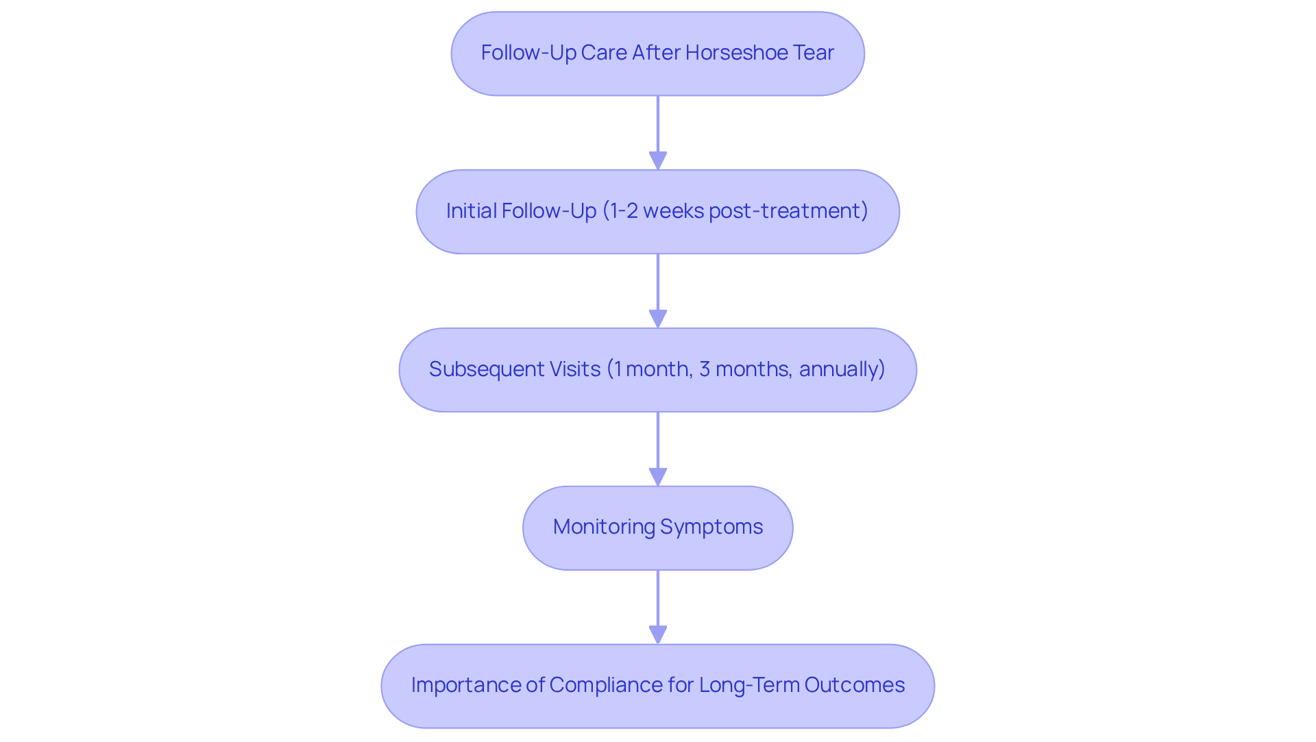
- Initial Follow-Up: A visit within one to two weeks post-treatment is crucial to assess healing and check for any new tears.
- Subsequent Visits: Additional check-ups are typically scheduled at one month, three months, and then annually, tailored to your individual risk factors and the severity of your initial injury.
- Monitoring Symptoms: It’s common to feel anxious about new symptoms, such as increased floaters or flashes of light. Please inform your eye care provider about these changes promptly, as new eye issues can arise weeks or months after the initial incident.
Statistics suggest that roughly 2.7% to 14% of treated eyes may develop new breaks in the eye tissue based on historical studies, with about 5% advancing to detachment (RRD). By keeping up with your follow-up appointments, you’re taking an important step in ensuring your retina stays healthy. A case study emphasized a patient who underwent multiple laser treatments for sequential retinal detachments and maintained 20/20 vision five years later, highlighting the effectiveness of timely follow-up care.
In Minnesota, compliance with follow-up care among patients with a horseshoe tear is critical. Studies show that adhering to follow-up schedules significantly impacts long-term outcomes. Remember, your commitment to follow-up care can make all the difference in protecting your vision for the future.
Conclusion
It’s understandable to feel concerned about your eye health, especially when faced with a horseshoe tear. This condition, marked by a U-shaped retinal injury, can lead to serious vision loss if left untreated. Recognizing symptoms like flashes of light, increased floaters, and blurred vision is crucial for early intervention. Remember, timely treatment options such as laser photocoagulation and cryopexy can effectively seal the tear and protect your vision.
We want to emphasize the importance of proactive care. Regular follow-up appointments are vital for monitoring your recovery and preventing further issues. By staying vigilant and seeking immediate medical attention when symptoms arise, you can significantly reduce the risk of complications like retinal detachment.
When you understand horseshoe tears, you can take steps to protect your eye health. Engaging with healthcare professionals and utilizing resources like the Be Eye Wise initiative can enhance your understanding and management of this condition. Taking these steps not only preserves your vision but also fosters peace of mind as you navigate eye health challenges. By prioritizing your eye health, you can ensure a brighter future for your vision.
Frequently Asked Questions
What is a horseshoe tear?
A horseshoe tear, also known as a flap lesion, is a specific type of retinal injury that appears U-shaped. It occurs when the vitreous gel pulls on the peripheral retina, often due to posterior vitreous separation (PVS).
Where do horseshoe tears typically occur in the retina?
Horseshoe tears typically occur at the back of the retina, particularly in the superotemporal and superonasal areas.
What are the potential complications of a horseshoe tear if left untreated?
If left untreated, horseshoe tears can lead to serious complications, including retinal separation, which poses a significant risk to vision.
What are the common symptoms associated with a horseshoe tear?
Common symptoms include floaters, flashes of light, and diminished vision, which may suggest the presence of a retinal rupture.
Who is most likely to experience a horseshoe tear?
Horseshoe tears are most commonly associated with middle-aged individuals, with no significant difference in occurrence between men and women.
What is the likelihood of advancing to retinal detachment if a horseshoe tear is symptomatic?
Symptomatic crescent-shaped disruptions have a 25 to 90 percent likelihood of advancing to detachment of the inner eye layer if not managed swiftly.
What should individuals with risk factors do regarding their eye health?
Individuals with risk factors such as high myopia or a history of lattice degeneration should have regular dilated eye examinations, even if they do not notice any symptoms.
What are some treatment options for horseshoe tears?
Treatment options include early detection and intervention methods such as laser photocoagulation or cryopexy, which can effectively seal the rupture and prevent further complications.
List of Sources
- Define Horseshoe Tear: Understanding the Condition
- Horseshoe Tear: Protecting Your Vision – Specialty Vision (https://specialty.vision/article/understanding-horseshoe-tears)
- Horseshoe or Flap Tear – EyeWiki (https://eyewiki.org/Horseshoe_or_Flap_Tear)
- Flap or Horseshoe Retinal Tear (https://optos.com/recognizing-pathology/definitions/flap-or-horseshoe-retinal-tear)
- Identify Symptoms: Recognizing Horseshoe Tear Indicators
- Horseshoe Tear: Protecting Your Vision – Specialty Vision (https://specialty.vision/article/understanding-horseshoe-tears)
- Horseshoe or Flap Tear – EyeWiki (https://eyewiki.org/Horseshoe_or_Flap_Tear)
- Retinal Breaks: Clinical Course and Outcomes After Retinopexy (https://retinalphysician.com/issues/2017/novdev/retinal-breaks-clinical-course-and-outcomes-after-retinopexy)
- Explore Treatments: Options for Managing Horseshoe Tears
- Other Treatments | Retina Associates of Utah (https://retinautah.com/retina-treatment/other-treatments)
- Retinal tear treatment: Options, recovery, and FAQs (https://medicalnewstoday.com/articles/retinal-tear-treatment)
- Horseshoe Tear: Protecting Your Vision – Specialty Vision (https://specialty.vision/article/understanding-horseshoe-tears)
- Cryopexy: Freezing Treatment for Retinal Tears – Specialty Vision (https://specialty.vision/article/cryopexy)
- Cryopexy: Freezing Treatment for Retinal Tears – NERA (https://retinamd.com/article/cryoretinopexy)
- Ensure Follow-Up Care: Monitoring and Long-Term Management
- Retinal Breaks: Clinical Course and Outcomes After Retinopexy (https://retinalphysician.com/issues/2017/novdev/retinal-breaks-clinical-course-and-outcomes-after-retinopexy)
- What to Expect After Retinal Tear Surgery | Retina Surgery Recovery Tips (https://retinala.com/what-to-expect-after-retinal-tear-surgery)
- The Importance Of Follow-Up Care After Eye Surgery | Retina Associates of Orange County (https://retinaorangecounty.com/2025/09/13/the-importance-of-follow-up-care-after-eye-surgery)
- Majority of Retinal Tears Post-PVD Develop After Six-Week Follow-up (https://reviewofoptometry.com/article/majority-of-retinal-tears-postpvd-develop-after-sixweek-followup)
- Surgery for Retinal Detachment: What to Expect at Home (https://myhealth.alberta.ca/Health/aftercareinformation/pages/conditions.aspx?hwid=abp2623)