Posted by: Northwest Eye in General on July 15, 2025
Overview
This article aims to help you understand the parts of the eye diagram and their functions, highlighting the importance of each component in the visual process. We recognize that many people may have questions about how their eyes work, and it’s perfectly normal to seek clarity. Structures like the cornea, lens, retina, iris, pupil, and optic nerve all work together to facilitate sight, and understanding these can be reassuring.
It’s common to feel concerned about eye health, especially with common conditions such as cataracts and diabetic retinopathy. We want to stress the necessity of patient education and routine examinations to maintain your eye health. Regular check-ups can make a significant difference, and we are here to help you through this process, ensuring you feel supported every step of the way.
Introduction
Understanding the intricate workings of the eye is essential for both medical professionals and patients alike. We understand that navigating eye health can feel overwhelming, and that’s why it’s important to know the parts of the eye. The diagram not only delineates the various structures—such as the cornea, lens, retina, and optic nerve—but also highlights their vital functions in the visual process. However, it’s common to feel uncertain about how these components interact and the implications for your vision.
Despite the widespread acknowledgment of eye health’s importance, many individuals remain unaware of these connections. How can a clearer understanding of eye anatomy empower you to take charge of your eye health and potentially prevent vision impairment? We are here to help you through this process.
Define the Parts of the Eye Diagram
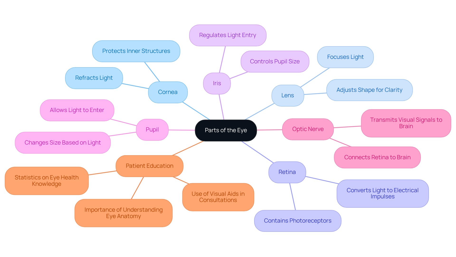
The parts of the eye diagram function as an essential visual aid that outlines the different structures of the eye and their connections. The parts of the eye diagram illustrate key components such as the cornea, lens, retina, iris, pupil, and optic nerve. Each structure plays a crucial role in the visual process, from concentrating illumination to transmitting visual signals to the brain. For instance, the cornea and lens work together to refract light, while the retina converts these light signals into electrical impulses for the optic nerve to relay to the brain.
We understand that comprehending this illustration is essential for both healthcare providers and patients. It not only aids in the accurate diagnosis and treatment of eye conditions, including cataracts, diabetic retinopathy, and dry eyes, but also empowers patients to take an active role in their eye health. Statistics reveal that over 80% of Americans consider themselves knowledgeable about eye health, yet many still lack a clear understanding of eye anatomy. This gap emphasizes the necessity for efficient educational materials, such as visual representations, to bridge knowledge gaps.
Real-world examples illustrate the effectiveness of eye anatomy visuals in patient education. During consultations, eye care professionals often utilize these diagrams to explain complex conditions like glaucoma or cataracts, enhancing patient comprehension and engagement. By visualizing the parts of the eye diagram, patients can better understand how specific conditions may impact their sight.
Ultimately, a is integral to effective eye care. As noted by health experts, “Eye health is integral to overall well-being, yet it is often overlooked until problems arise.” This underscores the importance of routine examinations and patient education in maintaining optimal eye health. If you are experiencing symptoms such as blurred sight, it is essential to seek professional medical help promptly. Furthermore, the annual cost of vision impairment in the United States is estimated at $134 billion, emphasizing the financial implications of neglecting eye health and the critical role of patient education in preventing costly outcomes.

Contextualize the Diagram in Eye Anatomy
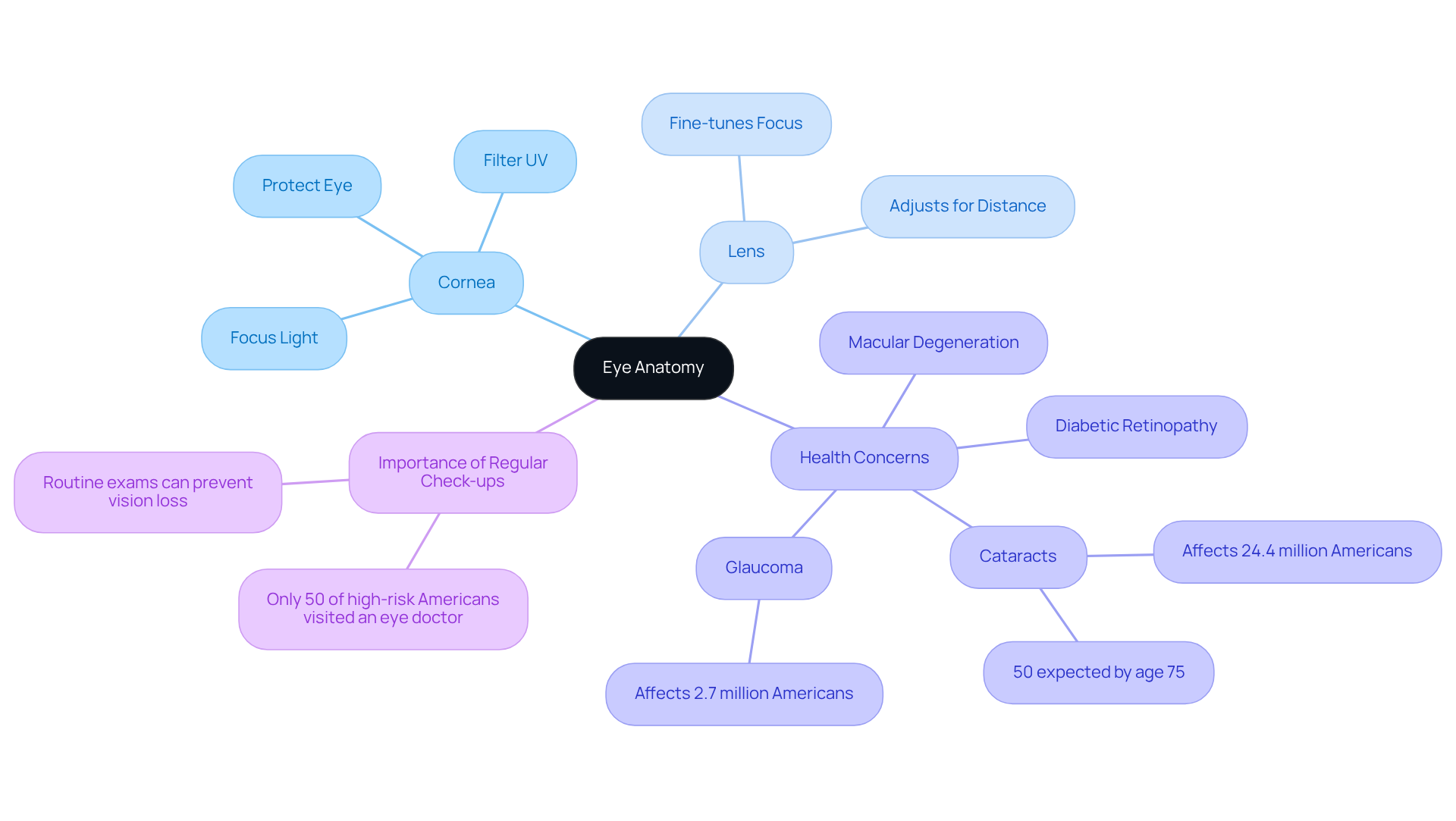
In the context of eye anatomy, the parts of the eye diagram serve as a foundational tool for understanding the complex operations of the eye as an organ. Each element, like the cornea, which bends rays and accounts for about 70% of the eye’s total focusing power, plays a specific role in the visual process. We understand that knowing how these components function can be reassuring. The cornea not only focuses light on the lens but also filters damaging UV light and protects the eye from germs and bacteria, highlighting its critical functions in maintaining overall eye health.
The diagram illustrates how the parts of the eye diagram interact, emphasizing the importance of maintaining eye health to ensure optimal function. It’s common to feel concerned about issues such as cataracts, which can influence the lens and result in unclear sight. This highlights the necessity for routine eye check-ups. Blurred sight may indicate the existence of eye conditions like cataracts, diabetic retinopathy, or macular degeneration, making it essential to seek professional diagnosis from a Northwest Eye doctor.
Statistics reveal that cataracts impact over 24.4 million Americans aged 40 and above, with nearly 50% expected to develop this condition by age 75. Additionally, the eye’s total optical power averages around 43 diopters, emphasizing its focusing capabilities. Understanding these components and their functions is crucial for . We are here to help you through this process, particularly given that over 90 million Americans are deemed high risk for loss or impairment of sight. Sight problems in this demographic have an annual economic impact of more than $145 billion.
Furthermore, advanced technologies like the Light Adjustable Lens (LAL) offer personalized vision correction options for cataract patients. This innovation sets a new benchmark in post-surgery outcomes, giving hope for better vision and quality of life.

Trace the Historical Development of Eye Diagrams
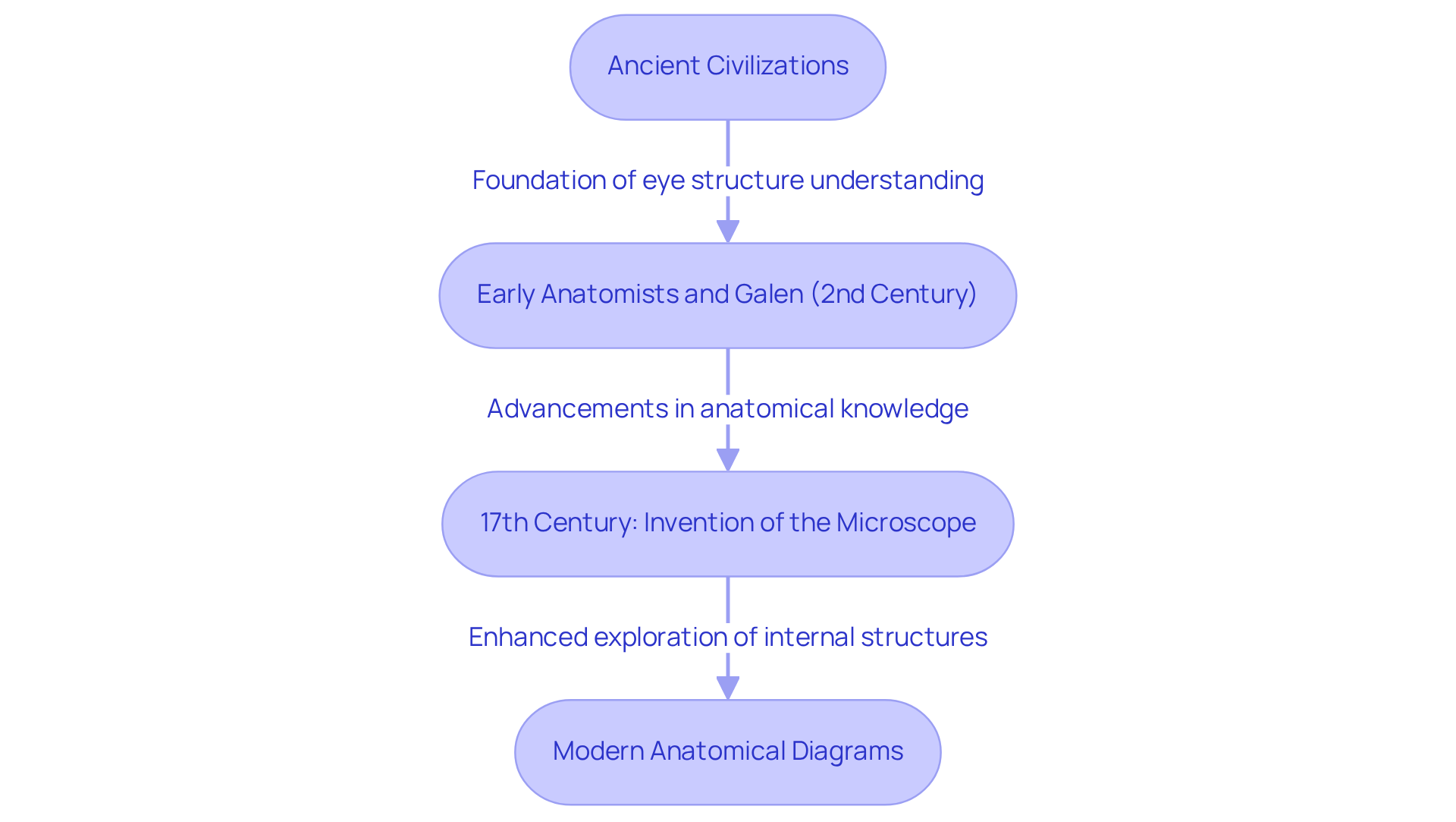
The historical evolution of eye representations is a fascinating journey that can be traced back to ancient civilizations. Early anatomists, motivated by a desire to understand the structure of the eye, laid the groundwork for what we know today. Notable contributions, such as the work of Galen in the 2nd century, provided some of the earliest descriptions of eye anatomy.
As time progressed, advancements in technology and medical understanding have led to more precise and comprehensive illustrations. We understand that this evolution can feel overwhelming, but it reflects our ongoing commitment to improving eye health. The invention of the microscope in the 17th century was a significant milestone, allowing for a deeper exploration of the eye’s internal structures.
This paved the way for , particularly the parts of the eye diagram, which are invaluable tools used in both education and clinical practice today. It’s common to feel a mix of curiosity and concern about our eye health, but rest assured, these advancements are here to support you in understanding your vision better.

Identify Key Components and Their Functions
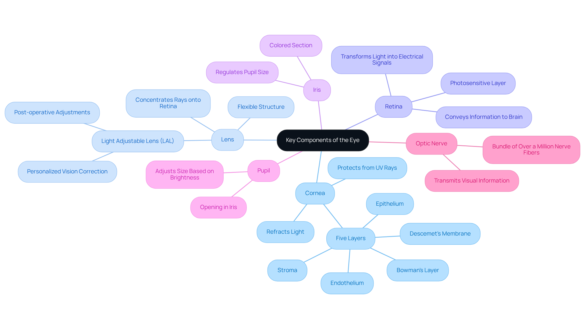
Key components of the eye include:
- Cornea: This transparent front layer refracts light entering the eye, contributing approximately 43 diopters to the eye’s total optical power. Composed of five layers—epithelium, Bowman’s layer, stroma, Descemet’s membrane, and endothelium—the cornea works diligently to maintain its clarity and refractive ability. It also plays a crucial role in safeguarding the eye from harmful UV rays, emphasizing its significance in overall eye health.
- Lens: A flexible structure that is among the parts of the eye diagram, further concentrating rays onto the retina and enabling clear sight at various distances. The innovative Light Adjustable Lens (LAL) offered at Northwest Eye represents a significant advancement in cataract surgery. This remarkable option allows for post-operative adjustments tailored to your lifestyle, thus providing a personalized approach to vision correction.
- Retina: The photosensitive layer illustrated in the parts of the eye diagram at the back of the eye transforms illumination into electrical signals, conveying visual information to the brain through the optic nerve.
- Iris: One of the parts of the eye diagram, the colored section that manages the size of the pupil, regulating the amount of brightness that enters.
- Pupil: The opening in the center of the iris, which is among the parts of the eye diagram, permits illumination to pass through and adjusts in size based on brightness conditions.
- Optic Nerve: It is one of the critical parts of the eye diagram, consisting of a bundle of over a million nerve fibers that transmits visual information from the retina to the brain, playing a crucial role in the visual process.
We understand that comprehending the roles of these components is crucial for identifying how different eye conditions, like cataracts, can affect sight. For example, cataracts can cloud the lens, obstructing light and resulting in blurred sight. As Dr. Myles Munroe stated, “Sight is a function of the eyes, but insight is a function of the heart.” By understanding the parts of the eye diagram, you can better appreciate the significance of treatments and interventions aimed at preserving or restoring your sight.
Additionally, it is noteworthy that approximately 33% of people in the United States experience astigmatism due to corneal distortion. This statistic highlights the . Innovative treatment options, such as the Light Adjustable Lens offered at Northwest Eye, further illustrate how understanding eye components can aid in effectively treating conditions like cataracts. We are here to help you through this process.

Conclusion
Understanding the intricate components of the eye is essential for anyone seeking to enhance their knowledge of eye health and anatomy. We recognize that navigating this information can feel overwhelming at times. The parts of the eye diagram serves as a vital educational tool, illustrating how each element, from the cornea to the optic nerve, contributes to the overall function of vision. This visual representation not only aids healthcare professionals in diagnosing and treating eye conditions but also empowers you, the patient, to take an active role in your eye care.
Key insights discussed throughout the article highlight the specific functions of each part of the eye. For instance, the cornea refracts light and protects the eye from harmful rays, while the retina converts light into electrical signals for the brain. It’s common to feel uncertain about these processes, but understanding them can foster a sense of control over your eye health. The historical context of eye diagrams underscores the evolution of our understanding of eye anatomy, showcasing the advancements that have led to more precise and comprehensive illustrations. This knowledge is crucial, especially given the prevalence of conditions like cataracts and diabetic retinopathy among the population.
Ultimately, a well-rounded comprehension of eye anatomy enhances individual awareness and emphasizes the importance of routine eye examinations and patient education. By taking proactive steps in understanding eye health, you can significantly reduce the risk of vision impairment and improve your overall quality of life. Engaging with educational resources and seeking professional advice when experiencing symptoms can lead to better outcomes. Remember, we are here to help you through this process and appreciate the remarkable complexity of the human eye.
Frequently Asked Questions
What is the purpose of the parts of the eye diagram?
The parts of the eye diagram serves as a visual aid that outlines the different structures of the eye and their connections, illustrating key components such as the cornea, lens, retina, iris, pupil, and optic nerve.
How do the cornea and lens function in the visual process?
The cornea and lens work together to refract light, which is essential for focusing images onto the retina.
What role does the retina play in vision?
The retina converts light signals into electrical impulses that are transmitted to the brain via the optic nerve.
Why is understanding the parts of the eye important for healthcare providers and patients?
Understanding the parts of the eye is crucial for accurate diagnosis and treatment of eye conditions, empowering patients to take an active role in their eye health.
What percentage of Americans feel knowledgeable about eye health?
Over 80% of Americans consider themselves knowledgeable about eye health, although many still lack a clear understanding of eye anatomy.
How do eye care professionals use eye anatomy visuals in patient education?
Eye care professionals utilize diagrams to explain complex conditions like glaucoma or cataracts during consultations, enhancing patient comprehension and engagement.
What is the financial impact of neglecting eye health in the United States?
The annual cost of vision impairment in the United States is estimated at $134 billion, highlighting the financial implications of neglecting eye health.
What should individuals do if they experience symptoms like blurred sight?
It is essential to seek professional medical help promptly if experiencing symptoms such as blurred sight.


