





Posted by: Northwest Eye in Eye Symptoms and Their Causes on August 30, 2026
Introduction
Noticing a difference in pupil size can stir up worry, leaving many to wonder what it means for their health. Anisocoria, the medical term for unequal pupil sizes, can arise from benign physiological variations or indicate more serious pathological conditions. It’s natural to feel anxious when noticing changes in your body, especially something as visible as pupil size. Recognizing the difference between harmless variations and serious conditions can empower you to seek the right care.
What should you do when faced with this common yet potentially alarming symptom? Understanding these differences can help you take the right steps toward ensuring your well-being.
Define Anisocoria: Understanding Unequal Pupil Sizes
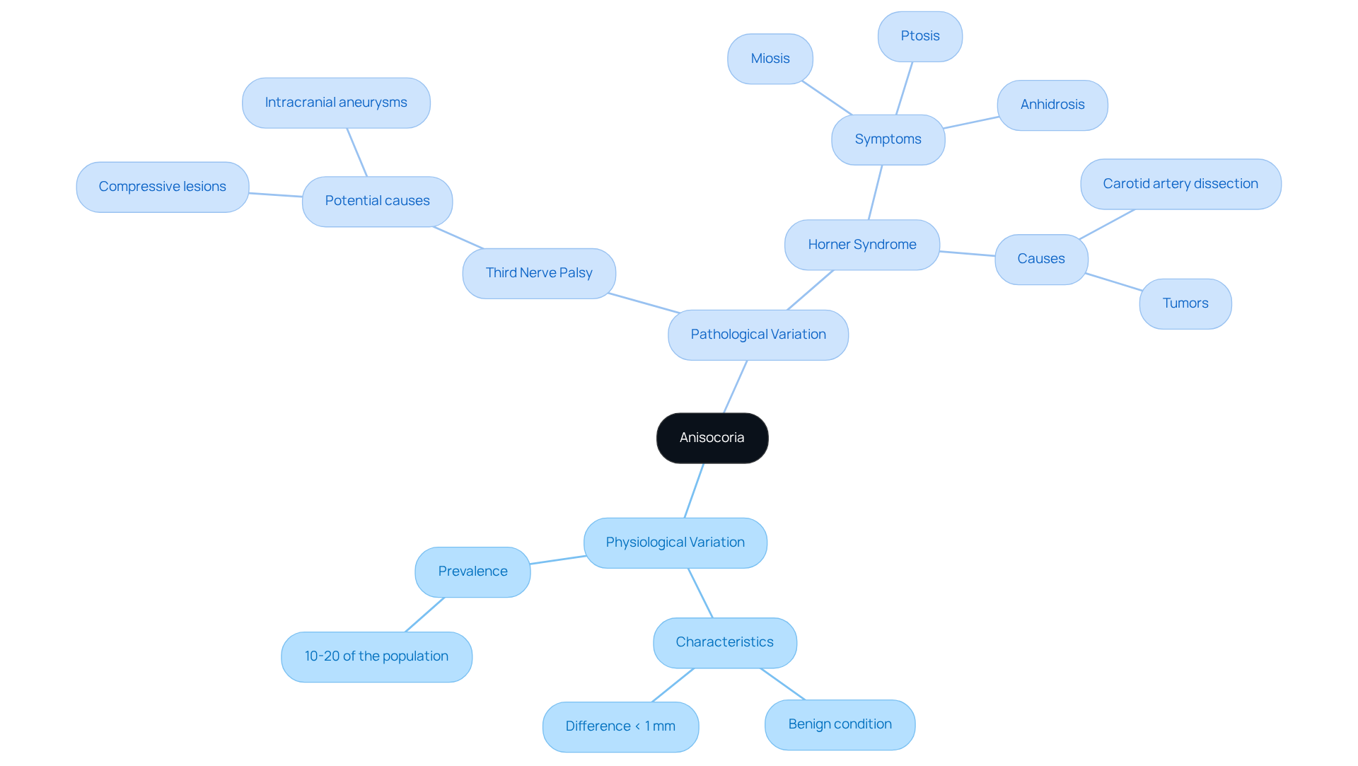
If you’ve noticed that you have different pupil size, you might be feeling concerned, and that’s completely normal. Anisocoria is when the openings of the eyes are unequal in size, also referred to as different pupil size, and it can be categorized into two types:
- Physiological variation, which is a benign difference seen in about 10 to 20% of the population.
- Pathological variation, which could understandably raise concerns about your health.
Typically, different pupil size is measured in millimeters; small discrepancies (less than 1 mm) are often harmless, while larger differences may necessitate further investigation.
Understanding different pupil size is essential, as it can indicate various neurological or ocular conditions, such as third nerve palsy or Horner syndrome. These conditions require immediate medical care, and seeking prompt attention can provide peace of mind and clarity. Recent studies suggest that up to 30% of individuals have a different pupil size, and it is normal for them.
We recommend a thorough clinical evaluation to help you understand your situation better, ensuring timely intervention when necessary. If you experience acute anisocoria, especially with other symptoms, it’s important to seek help right away. The interaction between the sympathetic and parasympathetic nervous systems regulates eye size, with the sympathetic pathway comprising a three-neuron arc originating in the hypothalamus. A comprehensive history and physical examination, including pupillary evaluation in different lighting scenarios, are essential for precise diagnosis and management. Remember, understanding your symptoms is the first step towards feeling better and regaining your peace of mind.
Explore Causes: Physiological vs. Pathological Anisocoria
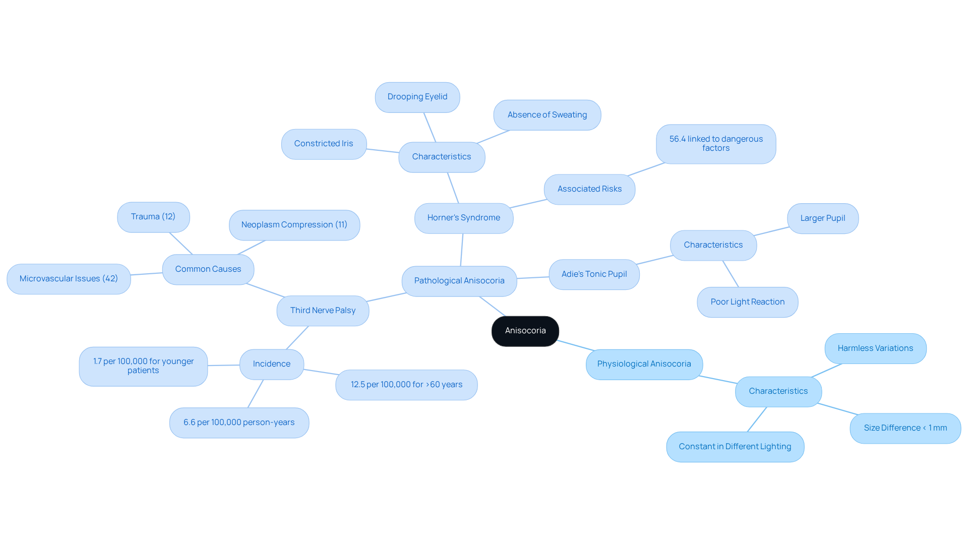
Noticing different pupil size can be unsettling, and it’s completely normal to have questions about what this means for your health. Anisocoria can be classified into two main categories: physiological and pathological. We want you to know that physiological differences in eye size are usually harmless and can be a normal part of how our bodies function. These variations are typically less than 1 mm and remain constant regardless of lighting. They often do not indicate any underlying health issues.
On the other hand, pathological unequal pupil size can arise from various medical issues, including nerve damage, eye trauma, or systemic diseases. Common causes of pathological anisocoria include:
- Third Nerve Palsy: This condition affects the oculomotor nerve, leading to pupil dilation and drooping eyelids. The annual incidence of acquired third-nerve palsy is approximately 6.6 per 100,000 person-years, with a significantly higher incidence in individuals over 60 years (12.5 per 100,000) compared to younger patients (1.7 per 100,000). The most prevalent factors contributing to third-nerve palsy include presumed microvascular issues (42%), trauma (12%), and compression from neoplasm (11%). If you experience this, neuroimaging is advised for all instances of acquired third-nerve palsy that do not have an apparent known origin.
- Horner’s Syndrome: This syndrome is characterized by a constricted iris, drooping eyelid, and absence of sweating on one side of the face. It can arise from various origins, including internal carotid artery dissection and tumors. A study revealed that 56.4% of Horner’s syndrome instances were associated with hazardous factors, highlighting the necessity for thorough assessment.
- Adie’s Tonic Pupil: This condition occurs when one pupil is larger and reacts poorly to light, often indicating underlying neurological issues.
Understanding these conditions can help you take the next steps towards ensuring your well-being and peace of mind, especially if you’re experiencing symptoms related to different pupil size.
Diagnose Anisocoria: Tests and Evaluations
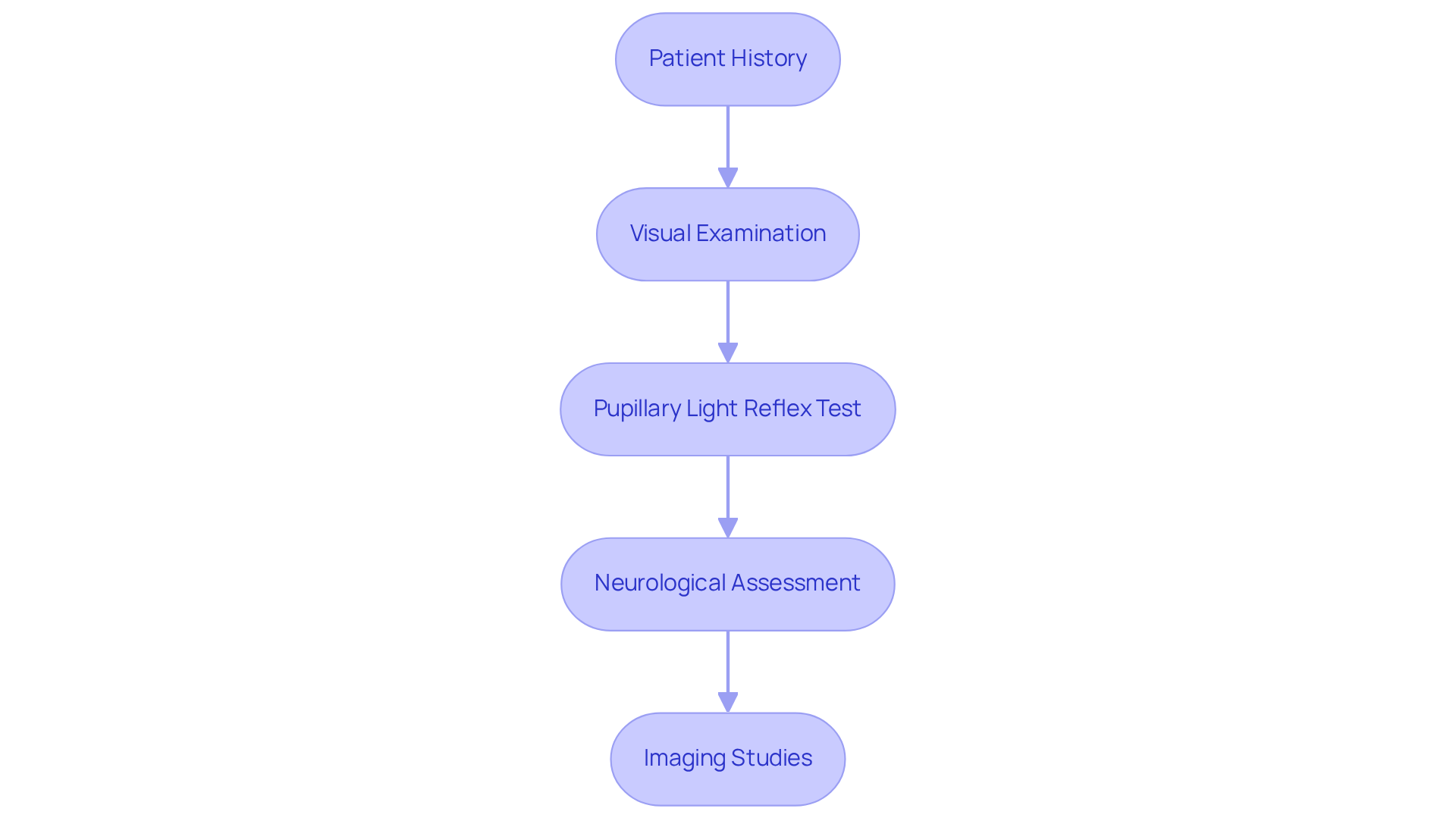
We understand that discovering different pupil sizes can be alarming, and it’s important to get the right care. Diagnosing anisocoria requires a comprehensive evaluation by an eye care specialist, typically involving the following steps:
- Patient History: Collecting detailed information about the onset of anisocoria, associated symptoms (like headaches or vision changes), and relevant medical history is crucial for understanding the context of the issue.
- Visual Examination: A thorough eye assessment evaluates iris size and reactivity to light in both bright and dim conditions, which is essential for identifying any abnormalities.
- Pupillary Light Reflex Test: This test evaluates how each pupil responds to light, helping to determine if one pupil is functioning abnormally, which can indicate underlying issues.
- Neurological Assessment: If necessary, a neurological examination may be conducted to exclude central nervous system issues that could lead to unequal pupil sizes.
- Imaging Studies: In cases where serious issues are suspected, imaging tests such as CT or MRI scans may be ordered to visualize the brain and surrounding structures, aiding in the diagnosis of potential life-threatening causes.
Did you know that about 10 to 20% of people have unequal pupil sizes? In most cases, this is harmless. However, non-physiological cases necessitate thorough examination and history-taking to identify underlying diseases. For example, it’s important to be aware that serious conditions, like a ruptured aneurysm, can be life-threatening, and early diagnosis is vital. A research analysis emphasized that different pupil sizes could signify serious issues such as acute brain injury, which may indicate elevated intracranial pressure or uncal herniation. It’s crucial to recognize that only 50% of patients survive a ruptured aneurysm after six months, which highlights the urgency of diagnosing serious issues related to different pupil sizes.
We believe that a thorough history and examination are key to understanding your unique situation. Furthermore, differential diagnosis is essential, as issues such as third nerve palsy or Horner syndrome may necessitate urgent intervention. Taking prompt action can make all the difference in ensuring your health and well-being.
Treat Anisocoria: Addressing Underlying Causes
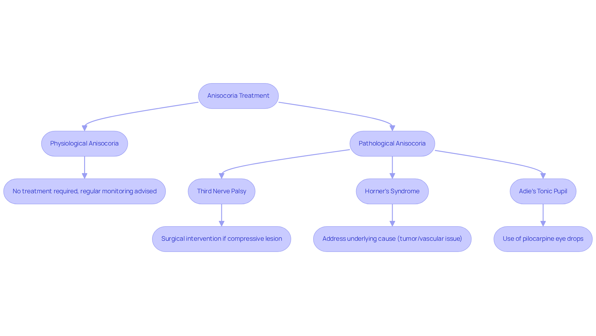
We understand that discovering anisocoria can be concerning and may raise questions about your health. Our focus is on understanding and addressing the underlying issues that contribute to anisocoria. Let’s explore some common treatment approaches based on the underlying causes, so you know what to expect:
- Physiological Anisocoria: Typically requires no treatment, as it is a benign condition. Regular monitoring may be advised.
- Pathological Anisocoria: Treatment varies depending on the underlying condition:
- Third Nerve Palsy: May require surgical intervention if caused by a compressive lesion.
- Horner’s Syndrome: Treatment focuses on the underlying cause, such as addressing a tumor or vascular issue.
- Adie’s Tonic Pupil: Management may include the use of pilocarpine eye drops to constrict the affected pupil.
In cases where anisocoria is linked to medication side effects, adjusting the dosage or switching medications may alleviate the condition. Rest assured, with the right diagnosis, effective management is within reach, allowing you to focus on your well-being.
Conclusion
We understand that noticing changes in your pupil size can be concerning, but it’s essential to know what these variations might mean for your health. Anisocoria means your pupils are different sizes. While this can be harmless, it might also point to more serious health issues. Differentiating between physiological and pathological anisocoria is crucial, as the latter may require immediate medical attention.
We want to emphasize how important it is to have a thorough evaluation. This includes:
- Looking at your medical history
- Conducting visual exams
- Conducting neurological exams
to accurately diagnose anisocoria. While many individuals may experience harmless variations, serious conditions like third nerve palsy or Horner’s syndrome necessitate prompt intervention.
By being aware of your pupil size differences, you can feel more confident in seeking medical care when it’s needed. Recognizing the signs and symptoms associated with anisocoria empowers you to take proactive steps towards your eye health, ensuring peace of mind and well-being.
Frequently Asked Questions
What is anisocoria?
Anisocoria is a condition characterized by unequal pupil sizes in the eyes, where one pupil is larger or smaller than the other.
What are the two types of anisocoria?
The two types of anisocoria are physiological variation, which is a benign difference seen in about 10 to 20% of the population, and pathological variation, which may indicate a health concern.
How is anisocoria measured?
Anisocoria is typically measured in millimeters, with small discrepancies (less than 1 mm) often being harmless, while larger differences may require further investigation.
What conditions can anisocoria indicate?
Anisocoria can indicate various neurological or ocular conditions, such as third nerve palsy or Horner syndrome, which require immediate medical attention.
How common is anisocoria among individuals?
Recent studies suggest that up to 30% of individuals may have a different pupil size, and it is considered normal for them.
What should I do if I experience acute anisocoria?
If you experience acute anisocoria, especially with other symptoms, it is important to seek medical help right away.
What factors regulate pupil size?
Pupil size is regulated by the interaction between the sympathetic and parasympathetic nervous systems, with the sympathetic pathway originating in the hypothalamus.
Why is a clinical evaluation important for anisocoria?
A thorough clinical evaluation, including pupillary assessment in different lighting scenarios, is essential for accurate diagnosis and management of anisocoria.
List of Sources
- Define Anisocoria: Understanding Unequal Pupil Sizes
- Anisocoria and Horner’s Syndrome – American Association for Pediatric Ophthalmology and Strabismus (https://aapos.org/glossary/anisocoria-and-horners-syndrome)
- Anisocoria: Background, Etiology, Pathophysiology (https://emedicine.medscape.com/article/1158571-overview)
- UMEM Educational Pearls | University of Maryland School of Medicine (https://medschool.umaryland.edu/em/cme/umem-educational-pearls?id=2617)
- Anisocoria – EyeWiki (https://eyewiki.org/Anisocoria)
- Anisocoria – StatPearls – NCBI Bookshelf (https://ncbi.nlm.nih.gov/books/NBK470384)
- Explore Causes: Physiological vs. Pathological Anisocoria
- Third Nerve Palsy (Oculomotor Nerve Palsy): Background, Etiology, Pathophysiology (https://emedicine.medscape.com/article/1198462-overview)
- Incidence and Causes of Acquired Third-Nerve Palsy: Population-Based Study (https://aao.org/eyenet/article/incidence-causes-of-acquired-third-nerve-palsy)
- Shedding light on Horner’s syndrome | Eye Care Network – Ophthalmology Times Europe (https://ophthalmologytimes.com/view/shedding-light-on-horner-s-syndrome)
- Understanding One Pupil Bigger Than The Other: Causes and Insights | Northwest Eye (https://nweyeclinic.com/understanding-one-pupil-bigger-than-the-other-causes-and-insights)
- Diagnose Anisocoria: Tests and Evaluations
- Anisocoria – StatPearls – NCBI Bookshelf (https://ncbi.nlm.nih.gov/books/NBK470384)
- A Clinical Approach to Pupil Testing | The Journal of Optometric Education (https://journal.opted.org/article/a-clinical-approach-to-pupil-testing-a-teaching-case-report)
- Anisocoria: Background, Etiology, Pathophysiology (https://emedicine.medscape.com/article/1158571-overview)
- Unequal pupils (https://www1.racgp.org.au/ajgp/2019/january–february/unequal-pupils)
- What Is Anisocoria? (https://aao.org/eye-health/diseases/what-is-anisocoria)
- Treat Anisocoria: Addressing Underlying Causes
- Cranial Nerve III Palsy (Oculomotor Palsy) – StatPearls – NCBI Bookshelf (https://ncbi.nlm.nih.gov/books/NBK526112)
- Diagnostic Considerations in an Acquired, Isolated Third Nerve Palsy (https://aao.org/eyenet/article/acquired-isolated-third-nerve-palsy)
- Self-Resolving Ischemic Third Nerve Palsy (https://webeye.ophth.uiowa.edu/eyeforum/cases/280-self-resolving-ischemic-TNP.htm)
- 3rd Nerve Palsy Surgery: Complex Eye Movement Restoration | The London Squint Clinic (https://londonsquintclinic.com/3rd-nerve-palsy-surgery-complex-eye-movement-restoration)
- Third Nerve Palsy (Oculomotor Nerve Palsy): Background, Etiology, Pathophysiology (https://emedicine.medscape.com/article/1198462-overview)