




Posted by: Northwest Eye in Glaucoma on October 17, 2026
Introduction
We understand that learning about optic nerve cupping can be concerning, especially when it occurs without glaucoma. This often-overlooked condition can signal underlying issues, and it’s important to address it to protect your vision. Together, we’ll explore the causes of optic nerve cupping, the symptoms to watch for, and the essential steps you can take for effective management. You might wonder what happens when the optic nerve changes shape and how you can take the right steps to protect your vision.
Define Optic Nerve Cupping and Its Importance
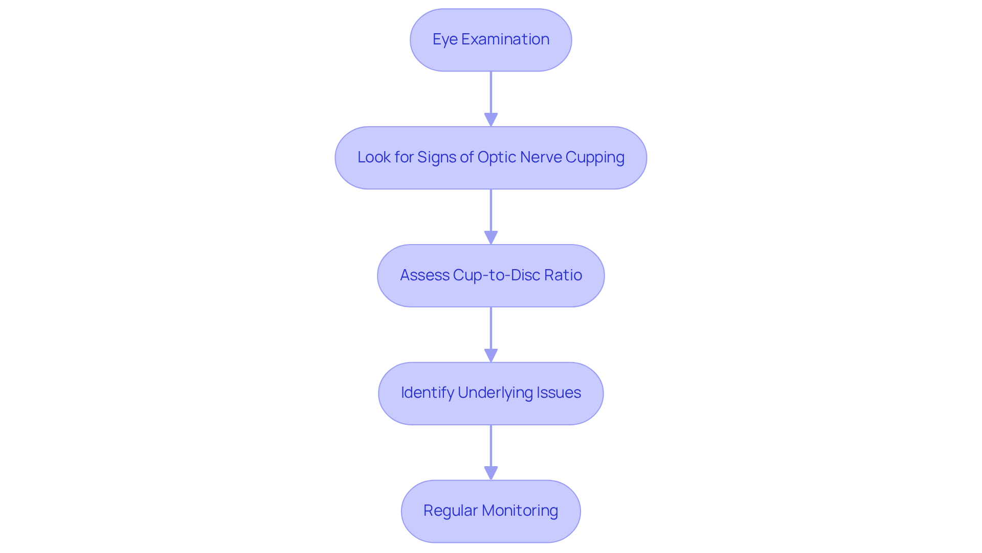
We understand that changes in your vision can be worrying, especially when it comes to your eye health. During a thorough eye examination, we look for signs like optic nerve cupping without glaucoma or excavation of the visual pathway head. This condition, characterized by optic nerve cupping without glaucoma, is defined by an increased cup-to-disc ratio, where the central cup of the visual pathway enlarges in relation to the surrounding disc. Comprehending these changes is essential, as they may indicate underlying problems impacting your sight, such as increased intraocular pressure or other diseases of the visual pathway.
It’s important to have regular eye exams, especially if you’re over 60 or have certain risk factors. Most adults should have comprehensive exams every one to two years, or annually after age 60. We want to ensure your eyes are healthy and your vision is clear. Early detection through regular eye exams can lead to effective management and peace of mind. Studies show that many patients retain excellent functional vision for decades when issues are identified and managed swiftly.
The assessment procedure generally encompasses a comprehensive array of tests utilized to evaluate visual pathway health. This includes sophisticated imaging methods such as Optical Coherence Tomography (OCT) and visual field analysis, which are essential for identifying optic nerve cupping without glaucoma as opposed to other sources of visual pathway damage. Progressive eye changes may result in the loss of peripheral vision, making early detection and monitoring essential to prevent irreversible vision impairment. By prioritizing your eye health, you can take proactive steps towards preserving your vision for years to come.
Explore Causes of Optic Nerve Cupping Beyond Glaucoma
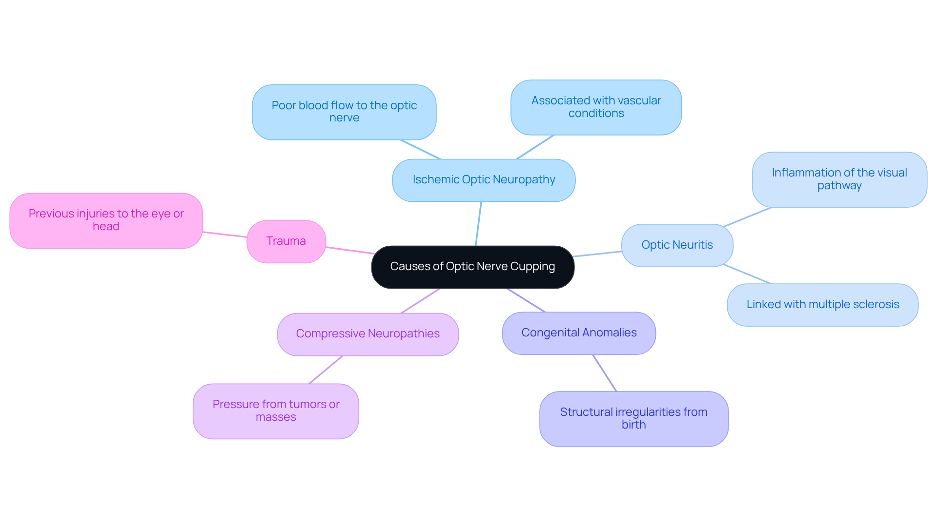
We understand that discovering the reasons behind eye disc deformation can be concerning, and it’s important to know that you’re not alone in this journey. While glaucoma is often the most recognized reason for this, there are several other conditions that can lead to similar changes:
- Ischemic Optic Neuropathy: Poor blood flow to the optic nerve can cause nerve fibers to die, leading to cupping.
- Optic Neuritis: Inflammation of the visual pathway can lead to depression of the disc, often linked with multiple sclerosis.
- Congenital Anomalies: Some individuals may be born with structural irregularities that predispose them to alterations in the visual pathway.
- Compressive Neuropathies: Tumors or other masses exerting pressure on the visual pathway can lead to deformation.
- Trauma: Previous injuries to the eye or head can lead to changes in the optic structure.
Understanding these factors is important for us as healthcare providers to distinguish between glaucomatous changes and optic nerve cupping without glaucoma. By recognizing these conditions, we can work together to ensure you receive the best possible care tailored to your needs.
Identify Symptoms of Optic Nerve Cupping
It’s concerning that many people may not realize they have a problem until it’s too late. Regular eye check-ups are essential for the early identification of optic disc changes, such as optic nerve cupping without glaucoma, which often occur without symptoms. As the condition progresses, you might notice several symptoms, including:
- Peripheral Vision Loss: Difficulty seeing objects outside of your direct line of sight, which can go unnoticed until significant damage occurs.
- Blurred Vision: A general decrease in visual clarity that may affect your daily activities. Blurred vision can result from various conditions, including cataracts, diabetic retinopathy, and uncorrected refractive errors, and should be evaluated by a Northwest Eye doctor.
- Difficulty with Night Vision: Increased challenges in low-light conditions, making it harder to navigate in dim environments.
- Visual Field Defects: Specific areas of vision loss detectable during comprehensive eye exams.
Recognizing these symptoms early can lead to timely diagnosis and treatment, potentially preventing further deterioration of your vision. We understand that individuals over 60 and those with certain risk factors may feel anxious about their eye health, but regular check-ups can provide peace of mind. If you notice any of these symptoms, we encourage you to reach out to a Northwest Eye doctor. We’re here to help you through this process. Remember, taking proactive steps today can help safeguard your vision for tomorrow.
Understand the Diagnostic Process for Optic Nerve Cupping
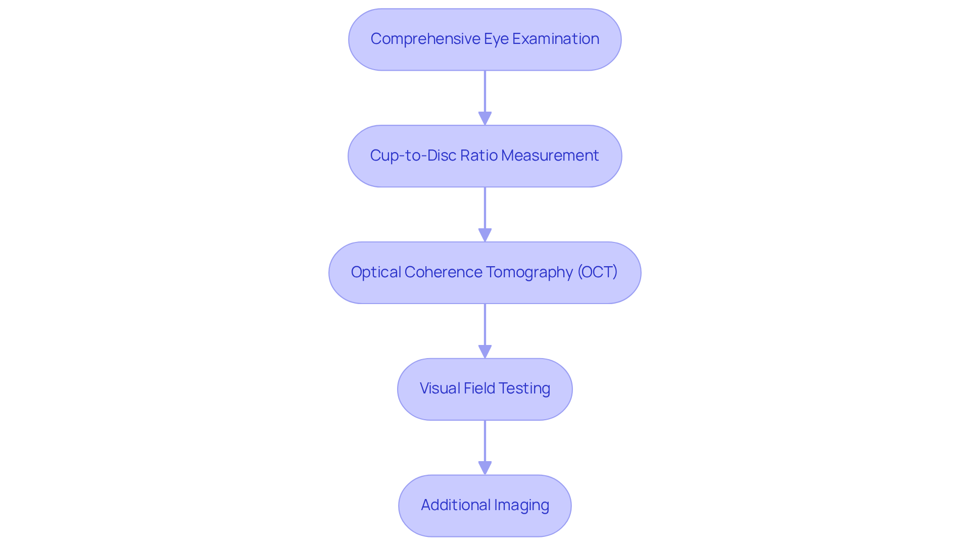
We understand that concerns about your vision can be overwhelming, especially regarding conditions such as optic nerve cupping without glaucoma. Here are the steps involved in the diagnostic process that can help ensure accurate assessment and management:
- Comprehensive Eye Examination: An eye care professional performs an extensive evaluation, including visual acuity tests and a detailed analysis of the visual pathway using an ophthalmoscope.
- Cup-to-Disc Ratio Measurement: The cup-to-disc ratio is measured, with a normal range typically between 0.3 to 0.4; a ratio less than 0.5 is generally considered normal.
- Optical Coherence Tomography (OCT): This advanced imaging method provides cross-sectional images of the visual pathway and retina, allowing for detailed analysis of fiber layers.
- Visual Field Testing: This assessment charts regions of vision impairment, assisting in the detection of any anomalies linked to damage of the visual pathway.
- Additional Imaging: In certain instances, further imaging examinations may be required to exclude other conditions that could resemble changes in the eye’s disc.
We encourage you to keep up with regular check-ups, as they play a vital role in monitoring your eye health and ensuring your treatment is on track, particularly if you are at risk for glaucoma or other ocular diseases. Adults should have annual eye exams after age 60 to ensure early detection of potential issues.
Recent advancements in diagnostic technology, such as machine learning algorithms, have shown an accuracy of 88 to 90 percent in identifying individuals with glaucoma, enhancing the diagnostic process. Moreover, research shows that the area under the curve (AUC) for distinguishing glaucomatous from non-glaucomatous optic nerve changes, such as optic nerve cupping without glaucoma, is remarkably high at 0.95, highlighting the effectiveness of these diagnostic methods.
Case studies emphasize the significance of assessing family history and physiological differences in the appearance of the visual pathways, which can inform further management and provide reassurance about the risk of developing glaucoma. Significantly, most individuals with disc changes do not experience blindness, and with early identification and intervention, you can feel confident about preserving your vision and living life to the fullest.
Manage Optic Nerve Cupping: Strategies and Treatments
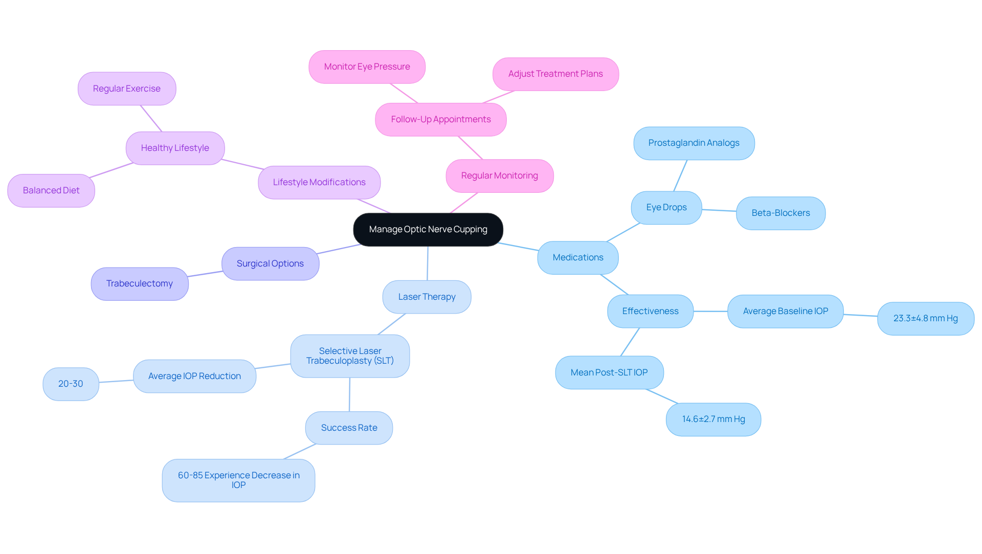
We understand that dealing with visual disc deformation can be a daunting experience, filled with uncertainty and concern for your vision. Management of this condition focuses on addressing underlying factors and preventing further harm. Let’s explore some effective strategies that can help you manage your condition and feel more secure in your eye health journey.
- Medications: Eye drops that lower intraocular pressure (IOP), such as prostaglandin analogs or beta-blockers, are commonly used to manage glaucoma-related cupping. Research shows that these medications can greatly lower IOP, with an average baseline IOP of 23.3±4.8 mm Hg and a mean post-SLT IOP of 14.6±2.7 mm Hg, which is vital for safeguarding the visual pathway.
- Laser Therapy: Selective laser trabeculoplasty (SLT) is a minimally invasive procedure that enhances fluid drainage from the eye, effectively lowering IOP. It’s common to feel apprehensive about procedures, but research shows that 60-85% of individuals experience a notable decrease in IOP after SLT, making it a viable option for those struggling with medication adherence.
- Surgical Options: In cases where medications and laser treatments are insufficient, surgical interventions such as trabeculectomy may be considered. This procedure establishes a new drainage route for fluid, assisting in reducing IOP and safeguarding the visual pathway.
- Lifestyle Modifications: Encouraging patients to maintain a healthy lifestyle, including regular exercise and a balanced diet, can improve overall eye health and circulation. These changes can support the effectiveness of medical treatments and enhance patient outcomes.
- Regular Monitoring: We know how important it is to stay connected with your eye care team. Ongoing follow-up with eye care professionals is crucial to observe changes in the visual pathway and modify treatment plans as needed. Regular follow-ups are essential to monitor eye pressure after laser treatment, ensuring that any advancement of optic nerve damage is addressed promptly.
By implementing these strategies, you can effectively manage optic nerve cupping without glaucoma and safeguard your vision. Taking these steps not only helps in managing your condition but also empowers you to take control of your eye health and future. Case studies demonstrate that patients who adhere to these management plans often experience improved outcomes, such as the finding that SLT alone achieved target IOP in 52.3% of eyes, highlighting the importance of a comprehensive approach to care. Additionally, expert insights indicate that “SLT is easy to perform, has an excellent safety profile, and is well tolerated by patients,” reinforcing the positive aspects of the treatment options discussed.
Conclusion
We know that concerns about your vision can be overwhelming, especially when faced with conditions like optic nerve cupping. This condition, which involves changes in the optic nerve’s structure, can arise from various causes beyond glaucoma, such as ischemic optic neuropathy, optic neuritis, and congenital anomalies. Recognizing the importance of regular eye examinations and being aware of the symptoms associated with optic nerve cupping can empower you to take proactive steps in managing your eye health.
Throughout this article, we’ve shared key insights regarding the diagnostic process, which includes:
- Comprehensive eye exams
- Advanced imaging techniques
- Visual field testing
These assessments are vital for distinguishing between glaucomatous and non-glaucomatous changes, ensuring that appropriate management strategies are implemented. Furthermore, effective treatment options such as:
- Medications
- Laser therapy
- Lifestyle modifications
were discussed, highlighting the importance of a tailored approach to care.
Ultimately, prioritizing your eye health through regular check-ups and being informed about conditions like optic nerve cupping can lead to better outcomes and preserved vision. By taking these steps, you can feel empowered and reassured about your eye health journey. We encourage you to engage with your eye care professionals, stay vigilant about any changes in your vision, and adopt healthy lifestyle practices. Your proactive attitude can make a significant difference in your long-term eye health.
Frequently Asked Questions
What is optic nerve cupping and why is it important?
Optic nerve cupping is characterized by an increased cup-to-disc ratio, where the central cup of the visual pathway enlarges in relation to the surrounding disc. It is important because it may indicate underlying problems affecting vision, such as increased intraocular pressure or other diseases of the visual pathway.
How often should I have my eyes examined for optic nerve cupping?
Most adults should have comprehensive eye exams every one to two years, or annually after age 60, especially if they have certain risk factors. Regular exams are crucial for early detection and effective management of eye health issues.
What tests are used to assess optic nerve cupping?
The assessment typically includes a variety of tests such as Optical Coherence Tomography (OCT) and visual field analysis, which are essential for identifying optic nerve cupping without glaucoma and distinguishing it from other sources of visual pathway damage.
What are some causes of optic nerve cupping beyond glaucoma?
Causes include ischemic optic neuropathy (poor blood flow to the optic nerve), optic neuritis (inflammation of the visual pathway), congenital anomalies (structural irregularities), compressive neuropathies (pressure from tumors or masses), and trauma (previous injuries to the eye or head).
Why is early detection of optic nerve cupping important?
Early detection is crucial because progressive eye changes can lead to the loss of peripheral vision. Identifying and monitoring these changes can help prevent irreversible vision impairment and ensure effective management of eye health.
List of Sources
- Define Optic Nerve Cupping and Its Importance
- Understanding Optic Nerve Cupping in Meriden, Connecticut – Meriden South Broad (https://meriden-sbroad.refocuseyedoctors.com/article/nerve-cupping)
- Optic Nerve Cupping (https://hospital.uillinois.edu/primary-and-specialty-care/ophthalmology/ophthalmology-services/optic-nerve-cupping)
- Explore Causes of Optic Nerve Cupping Beyond Glaucoma
- Anterior Ischemic Optic Neuropathy (AION): Background, Pathophysiology, Epidemiology (https://emedicine.medscape.com/article/1216891-overview)
- Ischemic Optic Neuropathy: Causes, Symptoms, and Treatment Insights | Northwest Eye (https://nweyeclinic.com/ischemic-optic-neuropathy-causes-symptoms-and-treatment-insights)
- Nonarteritic AION Incidence Increases After Age 50 (https://reviewofoptometry.com/article/nonarteritic-aion-incidence-increases-after-age-50)
- Identify Symptoms of Optic Nerve Cupping
- Optic disc cupping and glaucoma progression | Specsavers UK (https://specsavers.co.uk/eye-health/glaucoma/optic-disc-cupping-glaucoma-progression?srsltid=AU7gw4VAhDcIa2617HnUiG_tJfqS1Gif6z1Lb68IivfDOIFHWZH4hC3F)
- Optic Nerve Cupping – Specialty Vision (https://specialty.vision/article/understanding-optic-nerve-cupping-and-its-implications-for-eye-health)
- Optic Nerve Cupping: Protecting Your Vision from Glaucoma – Meriden-Curtis (https://meriden-curtis.refocuseyedoctors.com/article/optic-nerve-cupping)
- Understand the Diagnostic Process for Optic Nerve Cupping
- Understanding Optic Nerve Cupping: Causes, Diagnosis, and Care | Northwest Eye (https://nweyeclinic.com/understanding-optic-nerve-cupping-causes-diagnosis-and-care)
- Understanding Optic Nerve Cupping in Meriden, Connecticut – Meriden South Broad (https://meriden-sbroad.refocuseyedoctors.com/article/nerve-cupping)
- Evaluating and Managing Optic Disc Cupping in Children – Glaucoma Today (https://glaucomatoday.com/articles/2009-jan-feb/GT0109_06-php)
- Manage Optic Nerve Cupping: Strategies and Treatments
- Effectiveness of Selective Laser Trabeculoplasty as Initial or Adjunctive Treatment for Primary Open-Angle Glaucoma – PMC (https://pmc.ncbi.nlm.nih.gov/articles/PMC8784470)
- Understanding Laser Surgery for Glaucoma: Success Rates and Insights | Southwest Eye Care (https://southwesteyecare.net/understanding-laser-surgery-for-glaucoma-success-rates-and-insights)
- Laser Treatment for Glaucoma: A Breakthrough in Eye Care (https://albemarleeye.com/blog/laser-treatment-for-glaucoma)
- Understanding Optic Nerve Cupping: Causes, Diagnosis, and Care | Northwest Eye (https://nweyeclinic.com/understanding-optic-nerve-cupping-causes-diagnosis-and-care)