Posted by: Northwest Eye in General on August 30, 2025
Overview
Optical coherence tomography (OCT) is a vital non-invasive imaging technique that provides high-resolution, cross-sectional images of the eye. This technology is crucial for diagnosing conditions such as cataracts and retinal abnormalities. We understand that concerns about eye health can be overwhelming, and OCT plays an essential role in enhancing preoperative evaluations and surgical outcomes.
By detecting hidden retinal issues that standard examinations may miss, OCT significantly improves overall patient care and treatment planning. It’s common to feel anxious about potential eye conditions, but knowing that we have advanced tools like OCT can provide reassurance. We are here to help you through this process, ensuring you receive the best possible care.
Introduction
Optical coherence tomography (OCT) has emerged as a groundbreaking tool in modern ophthalmology, revolutionizing the way eye care professionals diagnose and treat various ocular conditions. We understand that navigating eye health can be daunting, especially for those facing cataract surgery. This non-invasive imaging technique offers unprecedented insights into the eye’s internal structures, particularly for cataract patients who may face hidden complications that could affect their surgical outcomes.
It’s common to feel uncertain about what this means for your care. However, with the rapid advancements in technology and the increasing reliance on OCT, one must wonder: how can patients ensure they are fully informed about the benefits and implications of this essential diagnostic tool? We are here to help you through this process.
Define Optical Coherence Tomography and Its Importance in Eye Care
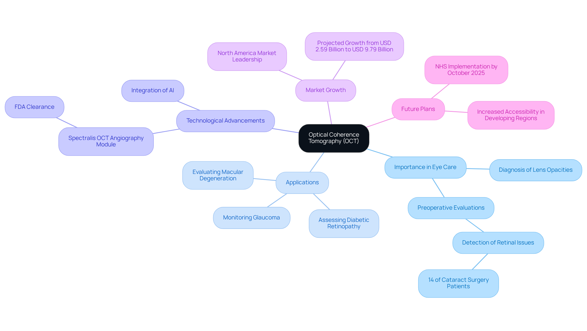
Optical coherence tomography (OCT) is an essential non-invasive imaging technique that employs light waves to generate high-resolution, cross-sectional images of the retina and various ocular structures. This advanced technology plays a crucial role in eye care, particularly in diagnosing and treating lens opacities. By providing a detailed view of the eye’s internal layers, OCT enables ophthalmologists to identify abnormalities that might be overlooked in traditional examinations, ultimately enhancing care management and treatment planning.
We understand that undergoing eye surgery can be a source of anxiety. The significance of optical coherence tomography (OCT) in lens surgery is underscored by its ability to uncover hidden retinal conditions that could impact surgical outcomes. A study found that 14% of individuals scheduled for cataract surgery had retinal issues detected by optical coherence tomography, despite standard fundoscopic examinations showing normal results. This finding highlights the into preoperative evaluations to ensure comprehensive care, especially when considering personalized lens options like the innovative Light Adjustable Lens (LAL) package available at Northwest Eye.
Recent advancements in optical coherence tomography technology, including the FDA-cleared Spectralis OCT angiography Module, have greatly enhanced imaging speed and quality, further solidifying its essential role in ophthalmology. The global optical coherence tomography market is projected to grow from USD 2.59 billion in 2025 to approximately USD 9.79 billion by 2034. This growth reflects the increasing prevalence of eye disorders and the rising demand for non-invasive diagnostic tools.
It’s common to feel uncertain about new technologies in healthcare. Statements from industry specialists emphasize the transformative impact of optical coherence tomography (OCT) on patient care. For example, the NHS in England plans to implement widespread OCT scans by October 2025, aiming to improve accessibility and reduce the need for hospital visits for eye care. Such initiatives illustrate a growing recognition of optical coherence tomography’s importance in enhancing diagnostic precision and individual outcomes in lens surgery, particularly when paired with advanced options like the LAL package that facilitates customized vision correction.
We are here to help you through this process, ensuring you feel informed and supported every step of the way.

Explain How Optical Coherence Tomography Works
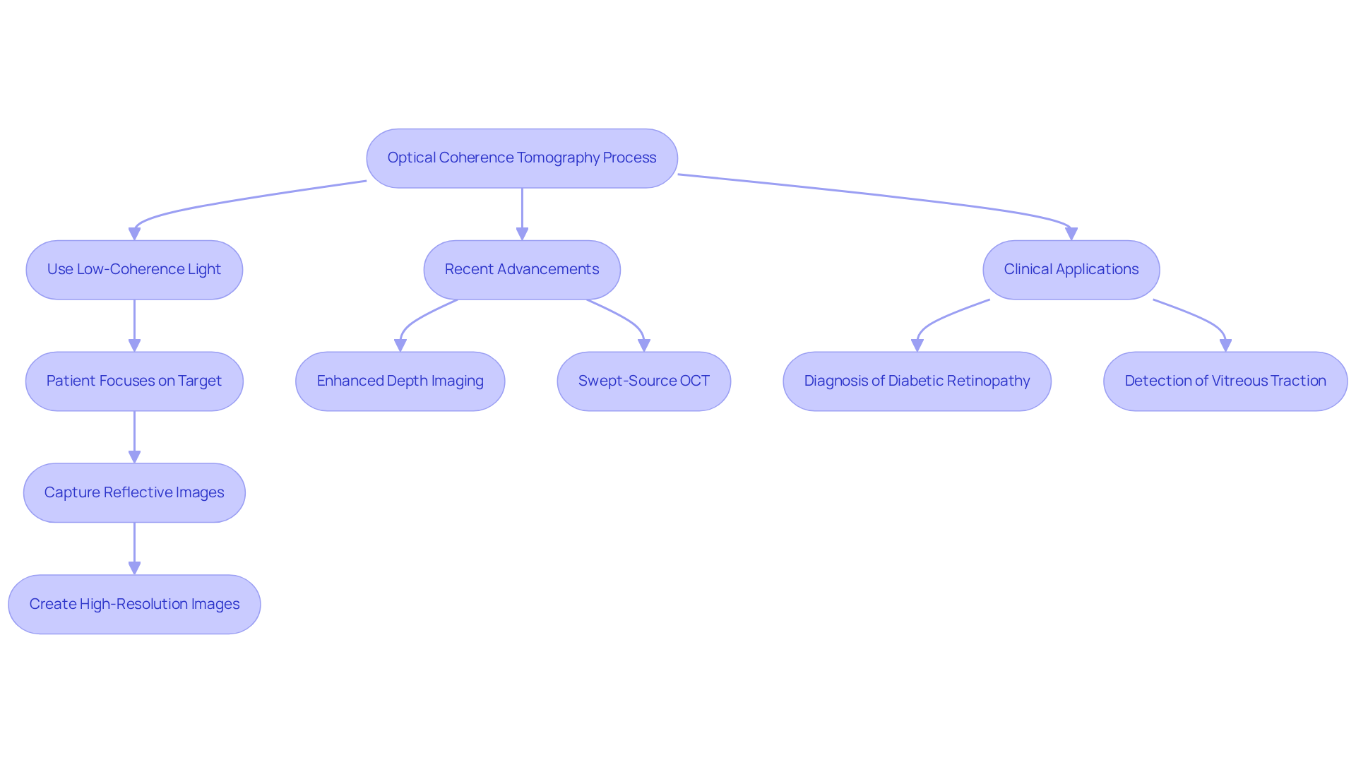
Optical coherence tomography (OCT) is a remarkable technology that uses low-coherence light waves to gently penetrate the eye and reflect off various internal structures, including the retina and optic nerve. This reflection is captured to create high-resolution, cross-sectional images of the eye’s anatomy. We understand that the use of a low-coherence light source is crucial, as it enhances imaging quality, allowing for . During the scanning process, patients focus on a designated target, enabling the OCT machine to capture images of each eye in just a few minutes. This non-invasive technique provides real-time data, which is invaluable for analyzing potential diseases or abnormalities.
Recent advancements in OCT technology have introduced innovative techniques, such as Enhanced Depth Imaging (EDI) and Swept-Source OCT (SS-OCT). These improvements not only enhance visualization of deeper retinal layers but also speed up the imaging process. We know how important these developments are in diagnosing conditions like diabetic retinopathy and glaucoma, where precise imaging is essential for effective treatment planning.
Case studies illustrate the efficacy of OCT in clinical settings. For instance, OCT has been instrumental in detecting vitreous traction, which can lead to retinal detachment. This highlights the importance of early diagnosis and intervention. Additionally, studies have shown that OCT can reveal nonzero autocorrelation in retinal images. This supports the hypothesis that cellular structures contribute to the observed intensity distributions, aiding in the early detection of retinal pathologies.
Overall, optical coherence tomography (OCT) serves as a fundamental element in contemporary ophthalmology. It equips eye care professionals with the resources essential for precise diagnosis and observation of various ocular conditions, including lens opacities. We are here to help you through this process, ensuring you receive the best possible care and support.

Discuss Applications of OCT in Diagnosing Eye Conditions
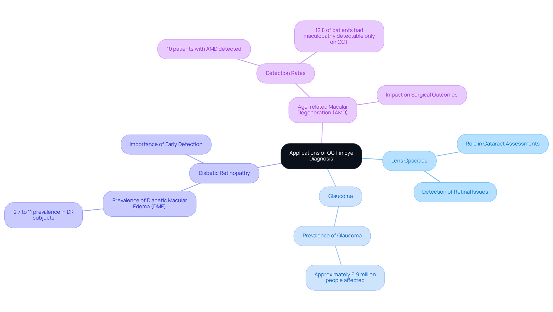
Optical coherence tomography (OCT) has become an essential tool for diagnosing various eye disorders, including lens opacities, glaucoma, diabetic retinopathy, and age-related macular degeneration (AMD). We understand that navigating eye health can be daunting, and optical coherence tomography is an that offers high-resolution cross-sectional images of the retina. This capability allows ophthalmologists to detect subtle structural changes that may indicate the onset of disease.
For individuals facing cataracts, OCT is particularly beneficial. It can uncover underlying retinal issues that might influence surgical outcomes. Recent studies reveal that optical coherence tomography (OCT) is capable of identifying conditions like epiretinal membranes and dry AMD, which often go unnoticed during standard examinations. In fact, a significant 14% of patients scheduled for lens surgery were found to have retinal conditions only detectable through optical coherence tomography. Among these, 80 individuals (12.8%) had maculopathy that standard evaluations did not identify.
Experts advocate for the routine use of optical coherence tomography in cataract assessments, emphasizing its role in enhancing care. By addressing potential complications before surgery, optical coherence tomography plays a crucial role in enhancing treatment success and patient satisfaction. We are here to help you through this process, ensuring that you receive the best possible care for your eyes.

Outline the Patient Experience During an OCT Test
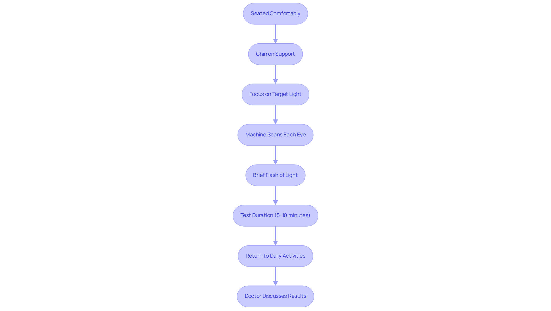
During an optical coherence tomography (OCT) test, we recognize that you may have concerns. You will be seated comfortably in front of the machine, resting your chin on a support to stabilize your head. It’s common to feel a bit anxious, but you will be instructed to focus on a target light while the machine scans each eye. This non-invasive procedure is quick, typically lasting between 5 to 10 minutes, and is designed to be painless. You might notice a brief flash of light during the scan; this is a normal part of the process and should not cause any discomfort.
After the test, you can immediately return to your daily activities. Your eye doctor will examine the results and with you, ensuring you feel informed and supported throughout this journey. Feedback from other patients highlights high satisfaction rates, with studies indicating that 83% of participants felt that optical coherence tomography improved their overall experience and reduced anxiety related to eye examinations. We are here to help you through this process, and we want you to feel reassured every step of the way.

Conclusion
Optical coherence tomography (OCT) represents a significant advancement in eye care, especially for those facing cataract surgery. By providing non-invasive, high-resolution images of the eye’s internal structures, OCT enhances the diagnostic process. This allows ophthalmologists to detect conditions that may otherwise go unnoticed, addressing your concerns about potential eye health issues. This technology not only aids in identifying lens opacities but also uncovers underlying retinal issues that could impact surgical outcomes, ultimately improving your care and treatment planning.
Throughout this discussion, we recognize the multifaceted role of OCT in ophthalmology. From its ability to identify subtle changes in the retina to its incorporation into preoperative evaluations, the importance of OCT cannot be overstated. Recent advancements, such as Enhanced Depth Imaging and Swept-Source OCT, further bolster its capabilities, ensuring that your eye care professionals are equipped with the best tools for accurate diagnoses. We want you to feel comfortable during OCT tests, as they are designed to be informative and alleviate anxiety, enhancing your overall satisfaction with eye care procedures.
As the demand for precise diagnostic tools continues to rise, integrating optical coherence tomography into routine eye examinations becomes crucial. We emphasize its role in improving patient outcomes, making it essential for both you and your healthcare providers to recognize and embrace this transformative technology. By prioritizing OCT in cataract assessments and other eye health evaluations, we can achieve a higher standard of care, ultimately contributing to better vision health for everyone. We are here to help you through this process, ensuring that you feel supported every step of the way.
Frequently Asked Questions
What is Optical Coherence Tomography (OCT)?
Optical coherence tomography (OCT) is a non-invasive imaging technique that uses light waves to create high-resolution, cross-sectional images of the retina and other ocular structures.
Why is OCT important in eye care?
OCT is crucial in eye care as it helps diagnose and treat lens opacities by providing detailed images of the eye’s internal layers, allowing ophthalmologists to identify abnormalities that may be missed in traditional examinations.
How does OCT contribute to lens surgery?
OCT plays a significant role in lens surgery by revealing hidden retinal conditions that could affect surgical outcomes. A study indicated that 14% of patients scheduled for cataract surgery had retinal issues detected by OCT, despite normal results from standard examinations.
What advancements have been made in OCT technology?
Recent advancements include the FDA-cleared Spectralis OCT angiography Module, which has improved imaging speed and quality, reinforcing the importance of OCT in ophthalmology.
What is the projected growth of the OCT market?
The global optical coherence tomography market is expected to grow from USD 2.59 billion in 2025 to approximately USD 9.79 billion by 2034, driven by the increasing prevalence of eye disorders and demand for non-invasive diagnostic tools.
How is OCT being implemented in healthcare systems?
The NHS in England plans to implement widespread OCT scans by October 2025 to improve accessibility and reduce hospital visits for eye care, highlighting the growing recognition of OCT’s role in enhancing diagnostic precision and outcomes.
What support is available for patients undergoing OCT?
Patients can expect support and information throughout the process to help them feel informed and comfortable with the use of OCT in their eye care and surgical planning.


